Imaging Contrast

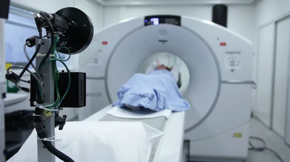
Contrast agents are injected into patients to help enhance images to make it easier for radiologists distinguish specific areas of the body from surrounding tissues. The most commonly used agents are iodinated contrast dye for computed tomography (CT), interventional cath lab angiography,  RF fluoroscopy, and in surgical OR procedures. MRI scans typically use gadolinium-based contrast agents. Ultrasound and echocardiography (cardiac ultrasound) imaging use contrast agents composed of microscopic bubbles to enhance images that otherwise would be suboptimal.

Thumbnail

Novel ‘double-contrast’ method spots small tumors on MRI

University of California, Davis, researchers believe their new probe could help detect very small, early stage tumors.

Thumbnail

University spinoff receives $750K to develop iron-based alternative to gadolinium contrasts

Ferric Contrast, a contrast agent company founded by faculty from the University at Buffalo in New York, says doctors and patients want new options in MR imaging.

Thumbnail

New MRI contrast agent detects cancers before they spread to the brain

The material increases magentic resonance sensitivity so that providers can visualize abnormal tissue changes indicative of metastasis.

Thumbnail

‘Game changer’: MRI contrast agent targets new liver cancer biomarker

Initial results were so successful that the material has been fast-tracked by the FDA in an effort to test its accuracy in human clinical trials.

Thumbnail

No gadolinium required: New take on old MRI contrast shows positive results

University of Texas at Dallas researchers applied their novel method to organic radical contrast agents with encouraging conclusions.

Thumbnail

ACR: Risk of administering CT contrast in patients with kidney disease ‘has been overstated’

The National Kidney Foundation joined the college in releasing a new consensus document that answers key questions and offers recommendations for using IV contrast in patients with impaired kidney function.

Thumbnail

‘One stop’ CT perfusion requires less contrast, radiation dose than CCTA

The new approach offers more comprehensive information with no loss of image quality, and may alter the care landscape for patients with coronary artery disease.

Thumbnail

Contrast-enhanced ultrasound predicts risk of progression to HCC

The researchers analyzed 39 consecutive patients with 44 pathologically confirmed cirrhotic nodules who first received a CEUS exam, followed by a traditional follow-up ultrasound every three or four months for their study.