Computed Tomography

Computed tomography (CT) is a fast and accurate imaging modality often used in emergency settings and trauma imaging. CT scans, with or without (or both) iodinated contrast are frequently used to image the brain, chest, abdomen and pelvis, but also have post-imaging reconstructive capabilities for detailed orthopedic imaging. It is now a standard imaging modality in emergency rooms to quickly assess patients. CT uses a series of X-ray images shot as the gantry rotates around the patient. Computer technology assembles these into into a dataset volume than can be slices on any access, or advanced visualization software can extract specific parts of the anatomy for study. Find more content specific to cardiac CT.

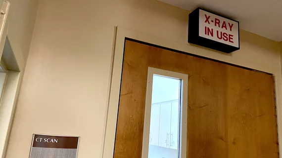
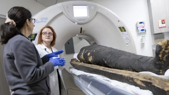
Paleo-radiologist: ‘I saw the king’s face for the first time’

Thanks to a radiologist equipped with CT and 3D image reconstruction, Pharaoh Amenhotep I is finally getting a proper clinical workup.

Thumbnail

Radiology trainees win over trauma surgeons with immediate reads

When trained with high-fidelity simulation, junior radiology residents can master the discipline of reading whole-body CTs right at the trauma scanner—and doing so with high diagnostic accuracy, work speed and interpretive confidence.

subtraction coronary CT angiography

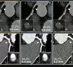
Subtraction boosts CCTA accuracy, even in the presence of extensive calcium

Researchers recently found results yielded via subtraction CCTA to be similar to those produced by ICA in assessing stenosis grading.

M&A mergers and acquisitions business deal

GE HealthCare eyes IR guidance company, releases revenue results

Less than a week after ringing the Nasdaq opening bell to showcase its long-awaited independence, GE HealthCare has announced a first strategic acquisition is in the works.

An example of virtual pulmonary valve implant planning software showing a virtual transcatheter pulmonary valve with measurements implnated in the virtual anatomy of a congenital heart patient. This was part of a training class at the Society of Cardiovascular Computed Tomography (SCCT) 2022 meeting.

FDA makes clearance easier for cardiac device implant simulation software

The FDA is reclassifying interventional cardiovascular implant simulation software into the class II (special controls) regulatory category, which will make it easier for the technology to pass regulatory approval. 

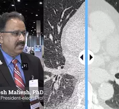
Mahadevappa Mahesh, PhD, professor of radiology and a medical physicist at the Johns Hopkins University School of Medicine, explains a new American College of Radiology (ACR) effort to ensure that lower radiation dose X-ray images under Image Wisely and As Low as Reasonable Achievable (ALARA) meet diagnostic reading standards. He spoke to Radiology Business at the Radiological Society of North America (RSNA) 2022 meeting.

VIDEO: Making sure lower dose X-ray is still diagnostic quality

Mahadevappa Mahesh, PhD, professor of radiology and a medical physicist at the Johns Hopkins University School of Medicine, explains a new ACR effort to ensure that lower radiation dose X-ray images under Image Wisely and As Low as Reasonable Achievable (ALARA) meet diagnostic reading standards.

Thumbnail

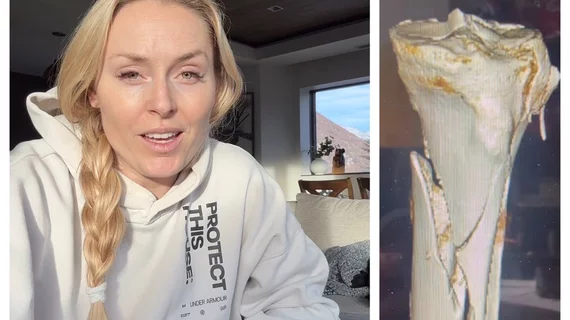
Bedside ultrasound found to effectively guide clinical decisions in trauma patients

Although whole-body CT scans are frequently one of the first assessments in the triage of trauma patients, new research argues that the use of bedside ultrasound could be more appropriate for initial examinations. 

A CT coronary calcium scoring exam at Northwestern Medicine's Central DuPage Hospital in the Chicago Suburbs. Dave Fornell

Trends in the number of radiology studies and a decline in radiation dose

A special report in Radiology offers a rare overview of the number of radiology exams performed each year worldwide and focuses on the decline of associated radiation doses patients receive.