Computed Tomography

Computed tomography (CT) is a fast and accurate imaging modality often used in emergency settings and trauma imaging. CT scans, with or without (or both) iodinated contrast are frequently used to image the brain, chest, abdomen and pelvis, but also have post-imaging reconstructive capabilities for detailed orthopedic imaging. It is now a standard imaging modality in emergency rooms to quickly assess patients. CT uses a series of X-ray images shot as the gantry rotates around the patient. Computer technology assembles these into into a dataset volume than can be slices on any access, or advanced visualization software can extract specific parts of the anatomy for study. Find more content specific to cardiac CT.

Internal surgery patients with infectious complications shown not to benefit by extra CT

Thoracic CT is safely and wisely omitted from diagnostic protocols for ICU patients who have signs of infection after abdominopelvic surgery but were already imaged with abdominal CT.

Homegrown tracking boosts follow-up imaging for incidental findings of uncertain gravity

A structured program to track incidental findings on body CT has significantly boosted rates of clinician follow-up as well as timely patient adherence to radiologist recommendations for next exams.

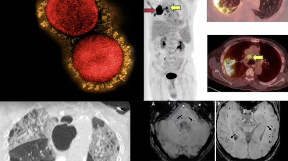
Appearances can be deceiving on chest CT performed for COVID in cancer patients

In a study of more than 250 COVID-positive patients with a history of any cancer, fewer than half the cohort had chest CT findings deemed typical for COVID-related pneumonia based on an RSNA classification guide. 

Thumbnail

Risk model-based lung cancer screening more cost-effective than USPSTF guidelines

Using risk model-based strategies to determine who should undergo low-dose CT lung cancer screening is more cost effective than current U.S. Preventive Services Task Force guidelines.

Q4 and full-year 2022 results in for GE HealthCare

GE HealthCare has some favorable tailwinds at its back as it proceeds into its first year unattached to a certain venerable parent corporation. 

AI helps reading-room radiologists differentiate colon cancer from diverticulitis

The model augmented and significantly improved diagnostic performance for abdominal subspecialists as well as residents—a result researchers say has major clinical implications.

Thumbnail

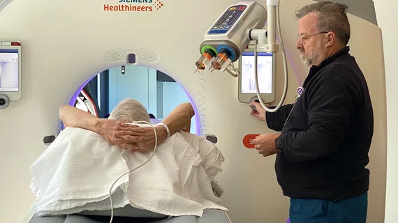
3D camera guidance meaningfully aids patient positioning, dose reduction

Regardless of a patient’s body mass index, using a scanner-integrated 3D camera for situating CT patients on the table can optimize not only patient positioning but also radiation dose. 

#CTA #acuteischemicstroke #AIS #radiomics

CTA-based radiomics can reliably estimate time since stroke onset

Radiomics features could be especially useful for determining TSS in scenarios where clinical data is lacking or unreliable, such as when patients have altered mental status.