Computed Tomography

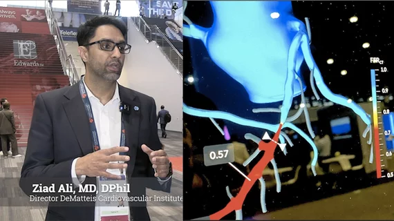
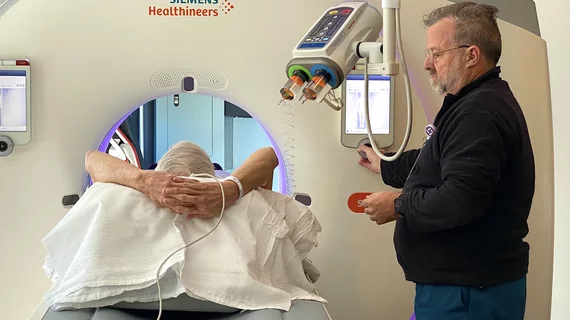
Computed tomography (CT) is a fast and accurate imaging modality often used in emergency settings and trauma imaging. CT scans, with or without (or both) iodinated contrast are frequently used to image the brain, chest, abdomen and pelvis, but also have post-imaging reconstructive capabilities for detailed orthopedic imaging. It is now a standard imaging modality in emergency rooms to quickly assess patients. CT uses a series of X-ray images shot as the gantry rotates around the patient. Computer technology assembles these into into a dataset volume than can be slices on any access, or advanced visualization software can extract specific parts of the anatomy for study. Find more content specific to cardiac CT.

Hip strain injury fracture broken hip

AI model predicts hip fractures in a short-term timeframe

Current risk assessments are unable to calculate the likelihood of a near-future fracture after a patient breaks their hip.

lung cancer screening

Patient-radiologist consultations, held in reading room after lung cancer screening, show promise

Conversations last 10 minutes or less, with specialists providing a preliminary interpretation of imaging findings in conjunction with smoking cessation counseling. 

Thumbnail

Study examines use of MSOT for detecting pancreatic cancer, improving surgery

The University of Oklahoma Health has earned a $3 million grant from the National Cancer Institute for this project.

Over the Shoulder Shot CT Brain Scan Images

Lower shift volumes lead to significantly fewer errors for neuroradiologists

When case volumes climb to 67-90 studies, error rates were 226% higher than those tackling 19 or fewer cases during their shift, experts reported. 

Medicare data stethoscope healthcare trends graphs finance financials

Use of CT angiography has surged in recent years, with a corresponding drop in MRI

Ease of access, lower cost, and shorter scan time, especially for emergent clinical indications such as acute stroke, all may be fueling CTA volumes. 

Thumbnail

FDA clears AI for breaking down lung CT images into subsegments

LungQ guides pulmonologists as they work to access peripheral locations within the lungs.

Thumbnail

System that makes brain and spinal surgeries more precise cleared by FDA

With the help of a CT or MR image, the SmartFrame OR and its ClearPointer optical navigation wand together provide guidance for instrument placement during neurological procedures.

chatgpt for patient questions about radiology

ChatGPT IDs incidental findings on CT images

New research suggests ChatGPT may be able to improve radiology workflows by performing an initial review of CT images.