Computed Tomography

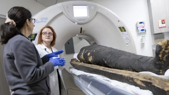
Computed tomography (CT) is a fast and accurate imaging modality often used in emergency settings and trauma imaging. CT scans, with or without (or both) iodinated contrast are frequently used to image the brain, chest, abdomen and pelvis, but also have post-imaging reconstructive capabilities for detailed orthopedic imaging. It is now a standard imaging modality in emergency rooms to quickly assess patients. CT uses a series of X-ray images shot as the gantry rotates around the patient. Computer technology assembles these into into a dataset volume than can be slices on any access, or advanced visualization software can extract specific parts of the anatomy for study. Find more content specific to cardiac CT.

Thumbnail

No need for updated imaging prior to major surgery in some cases, new data show

Prior chest imaging can sometimes be just as predictive of adverse events after surgery as updated imaging, according to new data set to be presented at the annual conference of the American College of Cardiology on March 5. 

Dual source CT scanners shown to expedite treatment in emergency settings

Dual source CT scanners can eliminate the need for beta blockers to control heart rates during coronary CTA exams.  

liver cancer

Pelvic CT after liver cancer: Costs versus benefits

Should patients treated for hepatocellular carcinoma undergo regular follow-up pelvic CTs? Rising survival rates may change the calculus.

Thumbnail

MRI catches CT in head-to-head lung imaging

When it comes to assessing patients with suspected pulmonary embolism, contrast-enhanced CT pulmonary angiography has no diagnostic edge over a certain free-breathing, unenhanced MRI perfusion protocol.

Thumbnail

Including radiologists in radiotherapy prep bolsters quality of care

Many radiation oncologists are not formerly trained in imaging interpretation, and radiologists’ collaborative participation in care planning can help to catch errors, experts wrote recently.

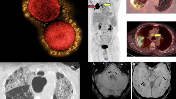
CT of coronavirus pneumonia, a solitary rounded ground-glass opacity (GGO) pattern. A 51-year-old woman in China presented in January 2020 without fever, but had close contact with positive patients. Top, baseline axial unenhanced chest CT obtained 6 days before the first positive PCR test. Bottom, chest CT scan 4 days later shows the size increase of the lesion (arrow). Image courtesy of RSNA. #COVID #SARSCoV2

How effective are chest CT severity scores in managing COVID?

While the studies on these systems have proven them to be effective in diagnosing and treating COVID in specific cohorts, the varying settings in which they were used can make it difficult to derive definitive conclusions on their efficacy.

Ed Nicol, MD, consultant cardiologist and honorary senior clinical lecturer with Kings College London and president-elect of the Society of Cardiovascular Computed Tomography (SCCT), explained artificial intelligence (AI) in cardiac CT is here to stay and its use is expanding. He noted that one AI-based algorithm is already included in recent cardiology guidelines and more will likely follow. #SCCT

Cardiac imagers need to understand AI as it enters clinical use and ACC guidelines

Most FDA-cleared AI algorithms are related to radiology and cardiology, meaning radiologists and cardiologists need to make an effort to learn how these technologies work.

Serial transverse CT scans in a 65-year-old man with severe COVID-19. Image courtesy of Radiology. #longcovid #covidlonghauler #lungdamagecovid

Severe COVID patients continue to evidence lung abnormalities 2 years after initial infection

Scans on these patients show a combination of fibrosis, thickening, honeycombing, cystic changes and dilation of the bronchi, among other imaging features.