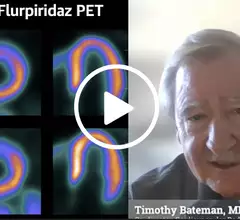
Videos

Example of a cancer that is difficult to see in dense breast tissue, but can be seen easier using 3D mammography digital breast tomosynthesis (DBT) breast imaging because the radiologist can go through the breast layer by layer if tissue..

VIDEO: The rapid adoption of 3D mammography and use of AI to address dense breasts

Stamatia Destounis, MD, a radiologist and managing partner at Elizabeth Wende Breast Care in Rochester, New York, chair of the American College of Radiology (ACR) Breast Commission, explains the rapid adoption of 3D mammogram digital breast tomosynthesis (DBT) technology.
 

Society of Breast Imaging (SBI) President John Lewin, MD, explains some of new initiatives and technology in mammography to increase earlier breast cancer detection. #SBI #breastimaging #mammography

VIDEO: SBI president outlines trends in breast imaging

Society of Breast Imaging President John Lewin, MD, explains some of the new initiatives and technology in mammography that are designed to increase early breast cancer detection.

VIDEO: Cardiac CT as a front-line chest pain exam in the ED

Harold Litt, MD, explains the use of cardiac CT as a primary imaging exam for chest pain at his emergency department over the past decade.

pacs.jpg

VIDEO: KLAS shares trends in enterprise imaging and AI

Monique Rasband, vice president of imaging, cardiology and oncology, KLAS Research, explains some of technology trends KLAS researchers have found in enterprise imaging system and radiology artificial intelligence (AI).

Charles E. Kahn, Jr., MD, MS, Editor of the the journal Radiology: Artificial Intelligence, and professor and vice chair of radiology at the University of Pennsylvania Perelman School of Medicine. He has been heavily involved in radiology informatics and has seen up close the evolution of radiology toward deeper integration with AI. #RSNA

VIDEO: Use cases and implementation strategies for radiology artificial intelligence

Charles Kahn, Jr., MD, editor of the the journal Radiology: Artificial Intelligence, and professor and vice chair of radiology at the University of Pennsylvania Perelman School of Medicine, explains the work involved integrating AI in radiology systems and the role of AI in augmenting patient care.
 

CT has been used for planning and procedural guidance in pulmonary vein isolation (PVI) for about 20 years. It shows the anatomy to the electrophysiologist. The EPs also can load the imaging into their EP mapping system. It also shows them the location of the esophagus and the phrenic nerve so they can avoid these during the procedures. #PVI #EPeeps

VIDEO: The expanding role of cardiac CT in electrophysiology

Harold Litt, MD, division chief of cardiothoracic imaging and director of the Center for Advanced CT Imaging Sciences at the Perelman School of Medicine of the University of Pennsylvania, explains the role of cardiac CT in EP procedures.

Charles E. Kahn, Jr., MD, MS, editor of the the RSNA journal Radiology: Artificial Intelligence, and professor and vice chair of radiology at the University of Pennsylvania Perelman School of Medicine. He discusses the need to validate artificial intelligence (AI) algorithms with your own patient population to determine if it is accurate for a specific institutions patients. He also explains how bias can be inadvertently added into a algorithm, and how the AI may take learning shortcuts. #AI

VIDEO: Assessing radiology AI and understanding programatic bias 

Charles E. Kahn, Jr., MD, MS, editor of the the RSNA  journal Radiology: Artificial Intelligence, and professor and vice chair of radiology at the University of Pennsylvania Perelman School of Medicine, discusses the need to validate AI algorithms with your own patient population data.  

VIDEO: Role of PFO closure in stroke prevention

John Carroll, MD, director of interventional cardiology and professor of medicine at the University of Colorado, explains the state of  transcatheter closure of patent foramen ovale (PFO) to prevent stroke.