Breast Imaging

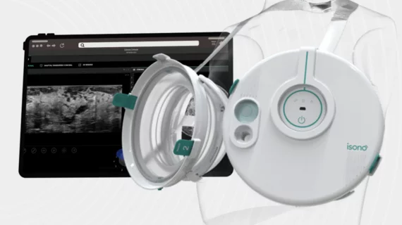
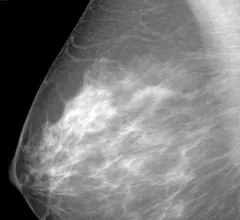
Breast imaging includes imaging modalities used for breast cancer screenings and planning therapy once cancer is detected. Mammography is the primary modality used. Mammogram technology is moving from 2D full-field digital mammography (FFDM) to breast tomosynthesis, or 3D mammography, which helps reduce false positive exams by allowing radiologists to look through the layers of tissue. Overlapping areas of dense breast tissue on 2D mammograms appear similar to cancers and 3D tomo helps determine if suspect areas are cancer or not. About 50% of women have dense breast tissue, which appears white on mammograms, the same as cancers, making diagnosis difficult. Radiologists use the Breast Imaging Reporting and Data System (BI-RADS) scoring system to define the density of breast tissue. Many states now require patients to be notified if they have dense breasts so they understand their mammograms might be suboptimal and they should use supplemental imaging that can see through the dense areas. This includes tomosythesis, breast ultrasound, automated breast ultrasound (ABUS), breast MRI, contrast enhanced mammography and nuclear imaging, including positron emission mammography (PEM).

COVID-19 medical imaging examples of various clinical presentations. SARS-CoV-2 clinical imaging presentations.

PHOTO GALLERY: What does COVID-19 look like on medical imaging?

This image gallery shows what the various clinical presentations associated with the COVID-19 virus that have been documented during the coronavirus pandemic.

Thumbnail

Research offers new guidance on managing architectural distortion visualized on DBT exams

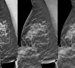
Multiple areas of architectural distortion visualized on digital breast tomosynthesis (DBT) exams are likely to produce high-risk pathology results. 

Thumbnail

The National Comprehensive Cancer Network has published new patient-friendly breast cancer screening guidelines

The new guidelines put emphasis on the age at which average-risk women should begin annual screening and breast assessments.

Thumbnail

Despite mandated coverage, nearly half of women are concerned about OOP breast cancer screening expenses

Women who skipped screening due to financial concerns outnumbered those who were not screened due to logistical issues such as scheduling and transportation conflicts, new survey data reveals. 

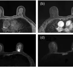
Example of various breast MRI protocol sequences that offer different types of soft tissue enhancement. Imaging performed on a Siemens Magnetom system. Breast MRI can help see through dense breast tissue to better detect cancers. #densebreasts #Breastdensity #BreastMRI

Abbreviated breast MRI protocols not as cost-effective as promised, new study shows

Experts found that although abbreviated protocols increased the amount of possible scans per hour from 4.7 to 18.8, non-scanning time rose from 50% to 74%.

Highly referenced breast density education hub makes patient-driven updates

Citing low health literacy as a barrier between providers and patients in overcoming healthcare disparities, DenseBreast-info.org updated their breast density materials to include more patient-friendly, “simple language.”

AI/radiologist combo improves breast cancer detection, decreases workloads

The new two-part AI system is based on a decision-referral approach and triages mammograms based on quantification of uncertainty.

Thumbnail

CEM vs breast MRI: Which is best for assessing treatment response?

A recent analysis of 51 patients revealed that contrast-enhanced mammography and MRI offered comparable assessments of lesion size, and both had similar specificity for pathologic complete response.