Breast Imaging

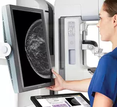
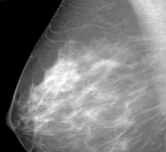
Breast imaging includes imaging modalities used for breast cancer screenings and planning therapy once cancer is detected. Mammography is the primary modality used. Mammogram technology is moving from 2D full-field digital mammography (FFDM) to breast tomosynthesis, or 3D mammography, which helps reduce false positive exams by allowing radiologists to look through the layers of tissue. Overlapping areas of dense breast tissue on 2D mammograms appear similar to cancers and 3D tomo helps determine if suspect areas are cancer or not. About 50% of women have dense breast tissue, which appears white on mammograms, the same as cancers, making diagnosis difficult. Radiologists use the Breast Imaging Reporting and Data System (BI-RADS) scoring system to define the density of breast tissue. Many states now require patients to be notified if they have dense breasts so they understand their mammograms might be suboptimal and they should use supplemental imaging that can see through the dense areas. This includes tomosythesis, breast ultrasound, automated breast ultrasound (ABUS), breast MRI, contrast enhanced mammography and nuclear imaging, including positron emission mammography (PEM).


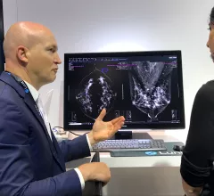
Supplemental breast imaging modalities of automated breast ultrasound (ABUS) and molecular imaging.

VIDEO: Use of supplemental imaging in women with dense breasts

Society of Breast Imaging (SBI) President John Lewin, MD, discusses the types of supplemental breast imaging used to aid cancer detection in women with dense breast tissue.

Two of the top stories this month included the death of MRI pioneer Raymond Vahan Damadian. Experts at MIT also developed ultrasound stickers that can be worn in the same manner as a Band-Aid while also producing diagnostic quality images in real-time. #MRI

Top Health Imaging stories in August 2022

Here are the most popular stories on Health Imaging in August, based on our website analytics. 

Synthetic MRI measures predict breast cancer treatment responses, according to new research

Researchers suggested that the quantitative measures derived from synthetic MRI exams could be used to help guide treatment decisions for breast cancer patients.

Surveillance breast MRI

Surveillance breast MRI findings linked with future second cancer

The new research focused specifically on associations between background parenchymal enhancement—a known risk factor for breast cancer, visualized on surveillance MRI exams—and secondary breast cancers.

breast cancer mammography mammogram

MR-directed enhanced mammography detects more malignancies than MR-directed ultrasound

MRI-directed contrast enhanced mammography could serve as a useful stand-alone or complimentary tool for biopsy planning when suspicious lesions are detected. 

Thumbnail

Minority women are more likely to delay or forego follow-up breast imaging, study reveals

Nearly 20% of the women included in the study did not complete their follow-up imaging at all, and out of the 3,648 women who did adhere to additional imaging, 23.3% delayed their exams beyond the recommended 60-day period.

Is stopping antithrombotic therapy prior to image-guide breast biopsy really necessary?

Experts arrived at this conclusion after comparing incidents of hematoma between patients who did and did not receive antithrombotic therapy prior to their procedure.

COVID's impact on breast radiology trainees represents a 'critical workforce issue'

The authors of the new study noted that, even before the pandemic, consultant breast radiologists were among the most in-demand specialists. The loss of interest in it as a specialty due to COVID will inevitably pose further challenges for recruitment.