Nuclear Medicine

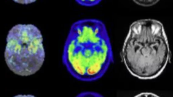
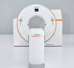
Nuclear medicine (also called molecular imaging) includes positron emission computed tomography (PET) and single photon emission computed tomography (SPECT) imaging. Nuclear imaging is achieved by injecting small amounts of radioactive material (radiopharmaceuticals) into patients before or during their scan. These can use sugars or chemical traits to bond to specific cells. The radioactive material is taken up by cells that consume the sugars. The radiation emitted from inside the body is detected by photon detectors outside the body. Computers take the data to assemble images of the radiation emissions. Nuclear images may appear fuzzy or ghostly rather than the sharper resolution from MRI and CT.  But, it provides metabolic information at a cellular level, showing if there are defects in the function of the heart, areas of very high metabolic activity associated with cancer cells, or areas of inflammation, data not available from other modalities. These noninvasive imaging exams are used to diagnose cancer, heart disease, Alzheimer’s and Parkinson’s disease, bone disorders and other disorders. 

Thumbnail

One chemistry professor's role in increasing radioisotope production in the U.S.

The work of a chemistry professor at the University of Wisconsin-Milwaukee is helping to blaze the trail of reducing the United States’ dependence on foreign countries for vital medical isotopes. 

PET/CT contrast debate: ‘Time to unleash the full power’ vs. ‘More is not always better’

Hybrid PET/CT enhanced with intravenous CT contrast deserves wider acceptance and adoption, as diagnostically optimized CT can complement PET—and vice versa—for a variety of potential indications. That’s one opinion on the matter.

Advnaces in nuclear cardiac imaging include the use of PET, quantitative coronary flow reserve and the additional CT ro SPECT and PERT scans. #ASNC

VIDEO: 2 key advances in cardiac nuclear imaging technology

Randall Thompson, MD, immediate past president of the American Society of Nuclear Cardiology (ASNC), explains two key advances in cardiac nuclear imaging.
 

New imaging biomarkers emerge for Alzheimer’s and its impairments

Brain MRI of the choroid plexus, the main supplier of cerebrospinal fluid, can deliver independent biomarkers for Alzheimer’s disease without an assist from clinical tests for amyloid abnormality or neurodegeneration. 

Thumbnail

New radiohybrid PET imaging agent increases detection of prostate cancer recurrence

For 45% to 47% of patients who were deemed negative after conventional baseline scans, the use of the radiohybrid imaging agent resulted in at least one true positive PET finding

A comparison on the black hole at the center of the Milky Way galaxy (left) with PET cardiac myocardial perfusion nuclear imaging exams (right).  The cardiac diagnosis of the galactic heart is motion artifact. Black hole image from the EHT Collaboration

Cardiologist and ASNC president diagnoses the heart of the galaxy based on black hole imaging

The first image of the black hole at the heart of our Milky Way galaxy amazed the scientific community, but left cardiologists with questions about the true cardiac health of the galaxy.

Thumbnail

PET/CT offers 'significant prognostic advantage' to patients newly diagnosed with multiple myeloma

The study, which followed 195 patients recently diagnosed with multiple myeloma (MM), found that a negative PET/CT scan six months after diagnosis and induction therapy was significantly linked with improved survival.

prostate cancer PSA

Total-body PET scanner detects cancer in 60 seconds

The new equipment, which initially will be used to treat prostate and neuroendocrine tumors, is expected to be operational in July.