Nuclear Medicine

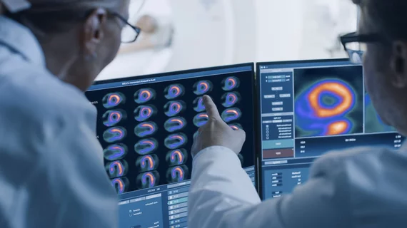
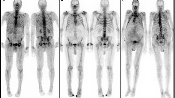
Nuclear medicine (also called molecular imaging) includes positron emission computed tomography (PET) and single photon emission computed tomography (SPECT) imaging. Nuclear imaging is achieved by injecting small amounts of radioactive material (radiopharmaceuticals) into patients before or during their scan. These can use sugars or chemical traits to bond to specific cells. The radioactive material is taken up by cells that consume the sugars. The radiation emitted from inside the body is detected by photon detectors outside the body. Computers take the data to assemble images of the radiation emissions. Nuclear images may appear fuzzy or ghostly rather than the sharper resolution from MRI and CT.  But, it provides metabolic information at a cellular level, showing if there are defects in the function of the heart, areas of very high metabolic activity associated with cancer cells, or areas of inflammation, data not available from other modalities. These noninvasive imaging exams are used to diagnose cancer, heart disease, Alzheimer’s and Parkinson’s disease, bone disorders and other disorders. 

Alex Ding, MD, radiologist and incoming AMA Board trustee, explains the new AMA policy that calls on Ciongress to change the requirements of the CMS requirement for appropriate use criteria clinical decision support software for all advanced imaging exams, including CT and MRI. #AMA #AMAmtg #AMA175 #AUC

VIDEO: AMA will ask Congress to change mandate on appropriate use criteria clinical decision support

Alexander Ding, MD, a radiologist and incoming American Medical Association (AMA) Board of Trustees member, explains the new AMA policy calling on Congress to change the language and implementation of the current Medicare mandate that all advanced medical imaging needs certification.

Researchers have designed a new cardiac SPECT imaging system that could potentially deliver images much faster than current models. The team presented its findings at the annual meeting of the Society of Nuclear Medicine and Molecular Imaging (SNMMI), SNMMI 2022.

The need for speed: New-look cardiac SPECT imaging system could be up to 100 times faster than current models

Researchers presented their proposed design of a self-collimating SPECT system at SNMMI 2022.

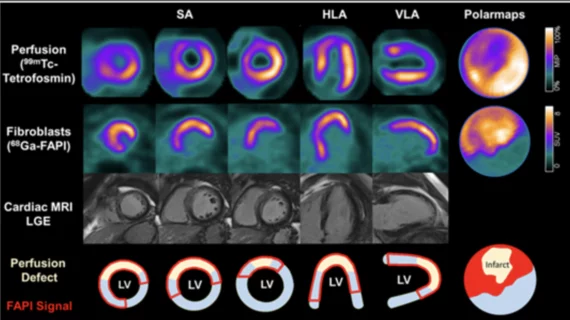
SNMMI Image of the Year 68Ga-FAPI-46 PET/CT heart attack acute myocardial infarction

‘Image of the Year’ highlights the predictive power of a new PET imaging agent

The Society of Nuclear Medicine and Molecular Imaging has chosen its 2022 Image of the Year, and it’s one that is sure to interest anyone in the field of cardiac imaging.

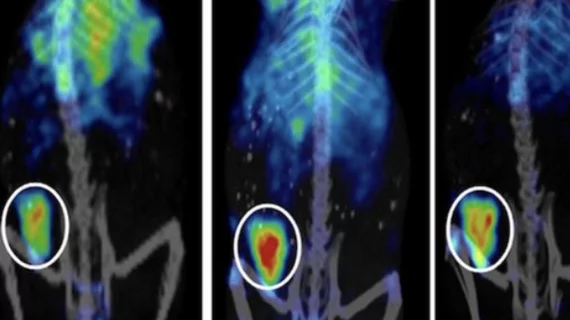
PSMA PET mapping improves radiation therapy contouring after PCA recurrence

Salvage radiation therapy guidelines for patients with prostate cancer recurrence need to be updated to include findings identified on PSMA PET, according to new research shared at SNMMI’s annual conference this week. 

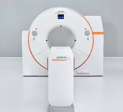
Siemens debuts new, cleared SPECT/CT model

Siemens Healthineers splashed an FDA-approved SPECT/CT system June 12 at the 2022 annual meeting of the Society of Nuclear Medicine & Nuclear Imaging in Vancouver, B.C.

Thumbnail

Cutting radiation exposure in half during PET/CT using virtual CT scans

“High-quality artificial intelligence-generated images preserve vital information from raw PET images without the additional radiation exposure from CT scans,” experts involved in the study explained.

NRC petitioned by patient advocacy group to require reporting of extravasation

The group is advocating for the NRC to treat significant extravasations in the same manner as any other reportable medical event. 

Thumbnail

Wisconsin company to begin producing medical isotopes next year

The company’s CEO, Greg Piefer, detailed the plans at the Wisconsin Entrepreneurs’ Conference on June 2, stating that production would be achieved via small-scale nuclear transmutation.