Nuclear Medicine

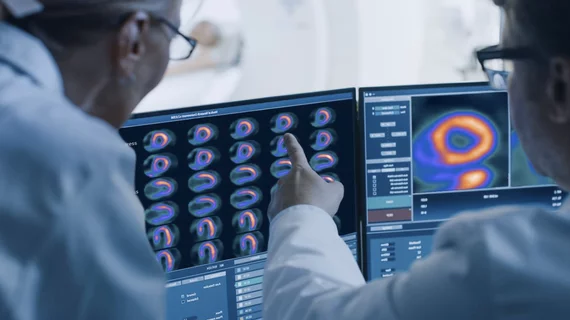
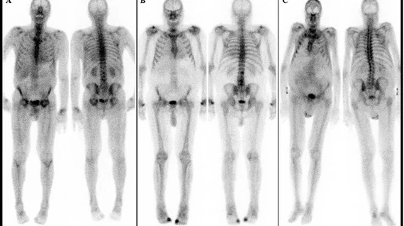
Nuclear medicine (also called molecular imaging) includes positron emission computed tomography (PET) and single photon emission computed tomography (SPECT) imaging. Nuclear imaging is achieved by injecting small amounts of radioactive material (radiopharmaceuticals) into patients before or during their scan. These can use sugars or chemical traits to bond to specific cells. The radioactive material is taken up by cells that consume the sugars. The radiation emitted from inside the body is detected by photon detectors outside the body. Computers take the data to assemble images of the radiation emissions. Nuclear images may appear fuzzy or ghostly rather than the sharper resolution from MRI and CT.  But, it provides metabolic information at a cellular level, showing if there are defects in the function of the heart, areas of very high metabolic activity associated with cancer cells, or areas of inflammation, data not available from other modalities. These noninvasive imaging exams are used to diagnose cancer, heart disease, Alzheimer’s and Parkinson’s disease, bone disorders and other disorders. 

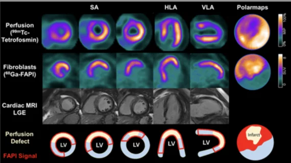
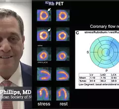
Comparison of flurpiridaz F-18 PET, SPECT and angiography of the 60-year-old female patients in the Aurora trial. The SPECT scan appeared normal, but flurpiridaz was shown to be more sensitive and showed the ischemia from two blockages in the right coronary artery. The new radiotracer may help expand cardiac PET. #ASNC

Flurpiridaz data shows promise to expand and enhance cardiac PET

The biggest news from the American Society of Nuclear Cardiology (ASNC) 2022 meeting was positive late-breaking data on the phase 3 Aurora trial for the flurpiridaz (F-18) PET radiotracer agent.

Thumbnail

FDG-PET shown to predict pancreatic cancer outcomes prior to surgery, could guide treatment decisions

FDG-PET scans in these patients allow clinicians to determine whether the tumors are still viable or not, thus playing a significant role in making treatment decisions.

Two of the top stories this month included the death of MRI pioneer Raymond Vahan Damadian. Experts at MIT also developed ultrasound stickers that can be worn in the same manner as a Band-Aid while also producing diagnostic quality images in real-time. #MRI

Top Health Imaging stories in August 2022

Here are the most popular stories on Health Imaging in August, based on our website analytics. 

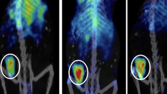
Two examples of PSMA-PET scans showing numerous prostate cancer metastases spread throughout the body. Many of these smaller tumors would not have been dected on previous standard-of-care imaging. Photo on left courtesy of SNMMI, right University of Chicago. #PSMAPET

PSMA-PET is rapidly changing the standard of care for prostate cancer patients

Adoption of PSMA-PET has been swift because it can significantly improve prostate cancer detection and treatment. SNMMI President Munir Ghesani, MD, explains how.

PET imaging uncovers a surprising new way COVID-19 affects the heart

A new study in JACC: Cardiovascular Imaging focuses on a potential new side effect of COVID-19, highlighting the continued importance of monitoring these patients going forward. 

5 ways to recruit sorely needed patients for Alzheimer’s clinical trials involving therapeutics

The demand is especially great for asymptomatic individuals of all races and ethnicities.

Whole-body PET image reveals body's immune response to COVID

In the study, patients who had recovered from an COVID infection were injected with a small amount of the 89Zr-labeled radiotracer before undergoing a uExplorer PET/CT scan.

COVID-19 medical imaging examples of various clinical presentations. SARS-CoV-2 clinical imaging presentations.

PHOTO GALLERY: What does COVID-19 look like on medical imaging?

This image gallery shows what the various clinical presentations associated with the COVID-19 virus that have been documented during the coronavirus pandemic.