Nuclear Medicine

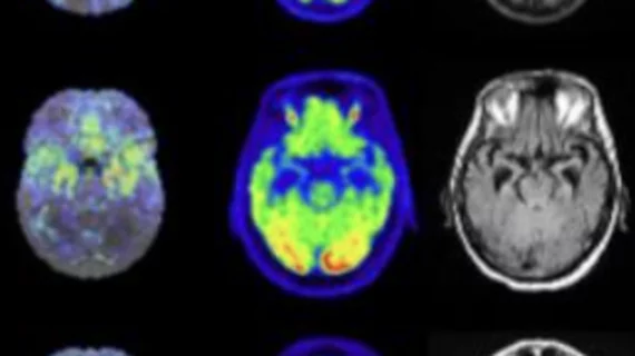
Nuclear medicine (also called molecular imaging) includes positron emission computed tomography (PET) and single photon emission computed tomography (SPECT) imaging. Nuclear imaging is achieved by injecting small amounts of radioactive material (radiopharmaceuticals) into patients before or during their scan. These can use sugars or chemical traits to bond to specific cells. The radioactive material is taken up by cells that consume the sugars. The radiation emitted from inside the body is detected by photon detectors outside the body. Computers take the data to assemble images of the radiation emissions. Nuclear images may appear fuzzy or ghostly rather than the sharper resolution from MRI and CT.  But, it provides metabolic information at a cellular level, showing if there are defects in the function of the heart, areas of very high metabolic activity associated with cancer cells, or areas of inflammation, data not available from other modalities. These noninvasive imaging exams are used to diagnose cancer, heart disease, Alzheimer’s and Parkinson’s disease, bone disorders and other disorders. 

Thumbnail

PET/CT findings predict post-treatment, radiation-induced hypothyroidism

Radiation-induced hypothyroidism is common yet underdiagnosed, potentially owing to a lack of follow-up consensus in patients treated with radiation therapy for head and neck cancers.

Thumbnail

FAPI PET/CT findings linked with risk of sudden cardiac death

Experts involved in the research suggested that these findings indicate a potential role for FAPI PET/CT imaging in detecting changes in myocardial fibrosis, noting that it could be more sensitive to earlier changes than standard cardiac MRI.

cerebral palsy PET imaging

How PET imaging could benefit children with cerebral palsy

These new findings could help providers differentiate and manage CP associated with different levels of motor impairment, authors of the study indicated.

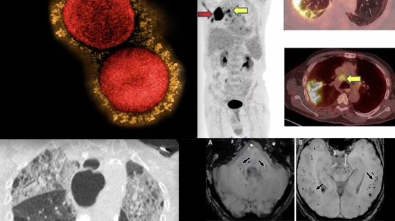
FAPI PET/CT shows promise as a better radiotracer for cancer than 18F-FDG. It also can be used for detection of infection and inflammation.

VIDEO: 6 key advances in molecular imaging technology for PET and SPECT

Munir Ghesani, MD, President of the Society of Nuclear Medicine and Molecular Imaging (SNMMI), system chief of nuclear medicine at Mount Sinai Health, explains recent advances in nuclear imaging technology.

Thumbnail

New lesion measurement assesses treatment responses more accurately than RECIST

Researchers believe the new method of assessing treatment responses could pave the way for developing new cancer therapeutics.

Thumbnail

A new Alzheimer's drug therapy appears effective. What might this mean for the future of amyloid PET?

Previously, CMS determined that coverage for patients receiving treatment was dependent on their being enrolled in a CMS-approved clinical trial under coverage with evidence development (CED).

A study published this week in the Journal of the American College of Cardiology (JACC): Cardiovascular Imaging shows artificial intelligence (AI) algorithms can more rapidly and objectively determine calcium scores in computed tomographic (CT) and positron emission tomographic (PET) images than physicians.[1] The AI also performed well when the images were obtained from very-low-radiation CT attenuation scans. https://doi.org/10.1016/j.jcmg.2022.06.006

Artificial intelligence can objectively determine cardiac calcium scores faster than doctors

A new study shows artificial intelligence (AI) algorithms can more rapidly and objectively determine calcium scores in CT and PET/CT images than physicians.

Thumbnail

Does the use of PSMA PET impact tumor upstaging?

A new risk analysis suggests the exam will increasingly result in tumor upstaging among high-risk patients.