Imaging Contrast

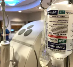
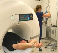
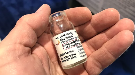
Contrast agents are injected into patients to help enhance images to make it easier for radiologists distinguish specific areas of the body from surrounding tissues. The most commonly used agents are iodinated contrast dye for computed tomography (CT), interventional cath lab angiography,  RF fluoroscopy, and in surgical OR procedures. MRI scans typically use gadolinium-based contrast agents. Ultrasound and echocardiography (cardiac ultrasound) imaging use contrast agents composed of microscopic bubbles to enhance images that otherwise would be suboptimal.

Test bolus of diluted contrast optimizes scan timing during CTA

It also reduces iodine uptake and radiation dose in patients, without sacrificing image quality.

Contrast protocols based on lean body weight save supplies, reduce patient risk

Adjusting contrast dose based on a patient’s lean body weight provides the same image quality as other administration methods, new data show.

The feasibility of reduced contrast flow rates in PE studies

Around one out of every 1,000 people in the U.S. are diagnosed with a pulmonary embolism every year, but even more undergo imaging to rule out clots.  

Thumbnail

Fluorinated contrast agents show 'strong potential' for MRI theranostics

Experts have developed a first-of-its-kind fluorinated contrast agent they believe will provide more detailed MR imaging and aid in the treatment of certain cancers. 

Thumbnail

GBCA doses can be reduced by over 50% for some MRI exams

In some cases, contrast doses can be reduced by as much as 62% without sacrificing diagnostic quality.

Dose reduction protocol makes multiple same-day contrast injections possible when necessary

This reduction protocol allows for acceptable lesion visualization while also providing a cautionary cushion when the safety of sequential contrast injections is in question.

Investigation into patient's death after contrast injection begins

The man's wife told the court that he had undergone similar scans multiple times before and had never experienced any sort of adverse reaction. 

time-delayed MRI phases improve brain tumor visualization

Using multiple MRI phases after contrast injection significantly improves visualization of brain tumors

Researchers are proposing making additional phases beyond the 10-minute mark mandatory when analyzing brain metastases.