Computed Tomography

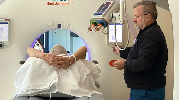
Computed tomography (CT) is a fast and accurate imaging modality often used in emergency settings and trauma imaging. CT scans, with or without (or both) iodinated contrast are frequently used to image the brain, chest, abdomen and pelvis, but also have post-imaging reconstructive capabilities for detailed orthopedic imaging. It is now a standard imaging modality in emergency rooms to quickly assess patients. CT uses a series of X-ray images shot as the gantry rotates around the patient. Computer technology assembles these into into a dataset volume than can be slices on any access, or advanced visualization software can extract specific parts of the anatomy for study. Find more content specific to cardiac CT.


Example of spectral, or dual-energy CT, confirming a pulmonary embolism (PE). Image courtesy of Philips Healthcare

Dual-energy CTA iodine maps offer small benefit in diagnosing pulmonary embolism

Dual-energy (DE) CT iodine maps offer a slight benefit when paired with traditional CT angiography images in diagnosing pulmonary embolism (PE), reported researchers in a Sept. 11 Radiology study.

Thumbnail

CCTA associated with reduced risk of death from coronary artery disease

Performing coronary CT angiography (CCTA) in addition to standard care is associated with a lower risk of death from coronary artery disease than standard care alone, according to research published in the New England Journal of Medicine.

Perivascular fat attenuation index CT scans showing the amount coronary wall inflammation. This measure is being tested in trials to see if it can accurately predict which coronary artery lesions will progress to cause heart attacks.

Novel cardiac imaging biomarker could predict risk of coronary inflammation, heart attack

A team of international researchers has developed a new imaging biomarker able to non-invasively predict a patient’s risk of coronary inflammation and heart attack, according to research published Aug. 28 in The Lancet.

Han awarded the inaugural 2018 DeHaan Award for Innovation in Cardiology

Vienna, VA (August 13, 2018) — B. Kelly Han, MD, is the recipient of the inaugural 2018 DeHaan Award for Innovation in Cardiology, which includes a $200,000 grant.

Thumbnail

Document offers guidance on curbing radiation exposure during CV imaging

An expert consensus document published May 2 in the Journal of the American College of Cardiology offers recommendations for limiting radiation exposure to both patients and medical personnel during cardiovascular imaging procedures.

Example of spectral, or dual-energy CT, confirming a pulmonary embolism (PE). Image courtesy of Philips Healthcare

MRA vs CTA for evaluating pulmonary embolism: Does the chosen modality impact downstream imaging utilization?

MR angiography (MRA) is a relatively new alternative to CT angiography (CTA) for the evaluation of suspected pulmonary embolism (PE) and it even has one major advantage over CTA: it does not expose patients to ionizing radiation.

Thumbnail

James K. Min named editor-in-chief of Journal of Cardiovascular Computed Tomography

James K. Min, MD, has been chosen to be the new editor-in-chief of the Journal of Cardiovascular Computed Tomography (JCCT), the official journal of the Society of Cardiovascular Computed Tomography (SCCT). He will serve a five-year term.

James Min named editor-in-chief of Journal of Cardiovascular Computed Tomography

Vienna, VA (April 25, 2017) — James K. Min, MD, FSCCT, has been selected for a five-year term as the new editor-in-chief of the Journal of Cardiovascular Computed Tomography (JCCT). He in turn has named Todd C. Villines, MD, FSCCT, as executive editor along with Gudrun Feuchtner, MD.