Computed Tomography

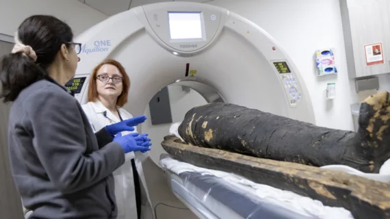
Computed tomography (CT) is a fast and accurate imaging modality often used in emergency settings and trauma imaging. CT scans, with or without (or both) iodinated contrast are frequently used to image the brain, chest, abdomen and pelvis, but also have post-imaging reconstructive capabilities for detailed orthopedic imaging. It is now a standard imaging modality in emergency rooms to quickly assess patients. CT uses a series of X-ray images shot as the gantry rotates around the patient. Computer technology assembles these into into a dataset volume than can be slices on any access, or advanced visualization software can extract specific parts of the anatomy for study. Find more content specific to cardiac CT.

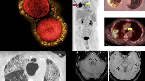
Imaging shows COVID vaccines effective at warding off pulmonary embolism

Researchers have found the condition significantly less among patients who received at least two doses of a COVID vaccine.

Stratifying patients by risk of poor outcomes could reduce overtreatment of lung cancer

Researchers are using radiomics to narrow patient cohorts down to those who are at the greatest risk of poor lung cancer outcomes.

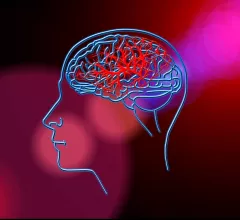
Ischemic stroke CT imaging. Images courtesy of RSNA

Stroke care still lags among certain Medicare populations

Despite increases of 250% for CTA and 428% for CTP, researchers observed lower rates of neuroimaging utilization among rural residents, older individuals (80 years and above), women and Black patients.

Thumbnail

AI spots pancreatic cancer in its earliest stages

Experts involved in the study suggest their findings could eventually be used to detect pancreatic cancer in its earliest stages when patients are most likely to respond to interventions favorably.

AI aids coma prognostics, potentially averting withdrawal of care

Deep learning has bested experienced neurosurgeons at predicting poor outcomes, including mortality, among patients admitted comatose with severe traumatic brain injuries.

Machine learning model quickly and accurately predicts outcomes for TBI patients

The model combines clinical data with imaging from head CT scans in individuals with severe traumatic brain injuries to quickly predict 6-month outcomes.

Ischemic stroke CT scan showing color coded blood flow for early and later arterial and venous contrast phases and areas of blocked blood flow. Image courtesy of RSNA

Disparities evident as CT stroke imaging rises sharply over 7-year period

Utilization of head CT perfusion (CTP) for imaging stroke patients spiked 428% between 2012 and 2019. Curiously, the uptrend was associated with higher mortality up to a year after discharge.

CT scans find prone position increases lung recruitment for COVID-19 patients

Using CT scans and EIT, researchers from an Italian hospital shed light on the specific benefits of the prone position for intubated COVID-19 patients.