Computed Tomography

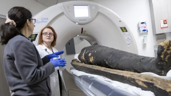
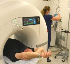
Computed tomography (CT) is a fast and accurate imaging modality often used in emergency settings and trauma imaging. CT scans, with or without (or both) iodinated contrast are frequently used to image the brain, chest, abdomen and pelvis, but also have post-imaging reconstructive capabilities for detailed orthopedic imaging. It is now a standard imaging modality in emergency rooms to quickly assess patients. CT uses a series of X-ray images shot as the gantry rotates around the patient. Computer technology assembles these into into a dataset volume than can be slices on any access, or advanced visualization software can extract specific parts of the anatomy for study. Find more content specific to cardiac CT.

Libertyville MRI

Smart shopping: Chicago orthopedic practice knows the value of a good service contract when investing in imaging equipment

Sponsored by Hitachi Healthcare Americas

The Illinois Bone and Joint Institute (IBJI) treats patients in the Chicago area for every orthopedic issue under the sun, from rheumatoid arthritis to total knee replacements, and its 100-plus physicians order thousands of imaging exams on an annual basis.

Thumbnail

How Secure Is That Scanner?

In a world of networked medical devices, it’s not hard to imagine a radiology-heavy cyberattack that is not only malicious but also ingenious.
 

Aidoc announces CE mark for the first AI-based workflow tool for Pulmonary Embolism

Aidoc’s solution for flagging and prioritizing pulmonary embolisms is now commercially available in Europe

Thumbnail

Plaque characteristics boost predictive power of CTA risk scoring

A CT angiography (CTA)-derived score that also incorporated the extent, location and composition of coronary plaque outperformed a model that focused only on the severity of stenosis, researchers reported Jan. 16 in JACC: Cardiovascular Imaging.

Thumbnail

SCCT shares updated guidance on CT use during TAVI, TAVR procedures

The Society of Cardiovascular Computed Tomography (SCCT) has issued an updated guidance for the use of CT in transcatheter aortic valve implantation (TAVI) and transcatheter aortic valve replacement (TAVR) procedures. The full document can be read in the Journal of Cardiovascular Computed Tomography.

Thumbnail

Industry group releases new guidelines for CT use in TAVR procedures

The Society of Cardiovascular Computed Tomography (SCCT) released updated guidelines for using CT imaging in patients undergoing transcatheter aortic valve replacement (TAVR).

Fujifilm announces entry Into U.S. computed tomography market with unveiling of FCT Embrace at ASTRO 2018

World's first 85cm 64 and 128 slice wide bore CT unit for oncology

FUJIFILM ANNOUNCES ENTRY INTO U.S. COMPUTED TOMOGRAPHY MARKET WITH UNVEILING OF FCT EMBRACE AT ASTRO 2018

STAMFORD, Conn.—FUJIFILM Medical Systems U.S.A., Inc., a leading provider of diagnostic imaging and medical informatics solutions, today introduced the FCT Embrace. Powered by Analogic, the FCT Embrace is the world’s first 85cm wide bore computed tomography (CT) imaging unitwith 64 or 128 slice configurations. Optimized for both oncology and radiology applications, the FCT Embrace, combined with other market-leading oncology solutions, offers enhanced and efficient CT Simulation with radiotherapy treatment planning capabilities. The unveiling at booth #3063 during the 2018 American Society for Radiation Oncology (ASTRO) Annual Meeting marks Fujifilm’s entry into the CT market, expanding its end-to-end diagnostic imaging product portfolio which is recognized for exceptional imaging at low dose.