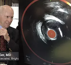
Researchers in Boston are set to co-lead a long-term study on the aging brain and factors that make individuals more vulnerable to neural injury and cognitive decline.
The team’s work will emphasize the role of specialized MRI techniques capable of detecting subtle changes around tumors with the help of pH-based imaging.
Using a virtual reality headset, the system, dubbed AR-VIU (augmented real-time volumetric imaging in ultrasound), creates a 3D rendering of anatomy based on 2D ultrasound images.
A team of experts recently developed the new system to differentiate between malignant and benign "second look" lesions on MRI for women with known breast cancer.
Those who undergo repeated exams before the age of 6 face almost double the risk of later developing intracranial tumors, leukemia or lymphoma, according to new data.
That’s according to an award-winning scientific online poster presented this week during the American Roentgen Ray Society’s annual meeting being held in Honolulu, Hawaii.
The hope is that the technique could lead to earlier detection of diseases by simultaneously identifying structural and functional abnormalities that standard ultrasound imaging methods alone cannot.
Features pertaining to location, density and superimposed structures were recently found to be associated with poorer outcomes for patients who initially had their lung cancer overlooked on radiographs.