Echocardiography

Cardiac ultrasound uses reflected sound waves (echos) to create images of anatomy inside the body. Echocardiograms are the primary cardiac imaging modality used to assess the heart and diagnose or track cardiac issues. Echo is the gold standard imaging modality to assess the heart, particularly with calculating left ventricular ejection fraction (LVEF), which is a measure of cardiac output. In addition to noninvasive standard transthoracic echo (TTE), invasive transesophgeal echo (TEE) is also used when clearer, more detailed imaging of the heart is needed. Both 3D and 4D echo echo systems are rapidly gaining wider adoption and enable new types of assessments, especially in the structural heart space and in transcatheter procedural guidance. Find news on general ultrasound imaging.

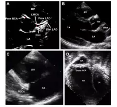
A figure from the ASE pediatric POCUS guidelines showing parasternal short-axis view demonstrating RV dilatation with bowing of the septum into the left ventricle, indicating pulmonary hypertension in this child with shock and pertussis.

New ASE guidance examines cardiac POCUS in children

The recommendations were developed by a writing group comprised of experts from different specialties, offering an array of perspectives and approaches.

March 2, 2023
candles funeral dead respect

Bijoy K. Khandheria, a cardiologist known for his generosity and kindness, dies at 66

"He will be deeply missed by everyone who had the pleasure of interacting with him and learning from him," according to a loving tribute from the American Society of Echocardiography.

February 15, 2023
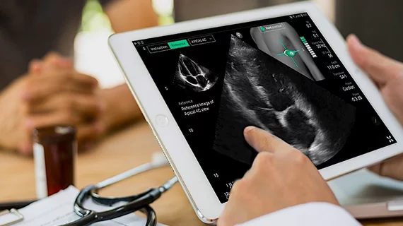
GE healthcare has purchased Caption Health. This added cardiac ultrasound AI guidenance to GE's echocardiogram systems so even inexperienced sonographers and point of care ultrasound (POCUS) users will be able to obtain high-quality echo exams.

GE Healthcare to acquire AI imaging specialists Caption Health

Caption Health has been a major player in the cardiac imaging space in recent years, gaining attention for its AI-powered echocardiography solutions. 

February 10, 2023
A 3D intracardiac echo (ICE) view of a surgical mitral valve using the NuVision ultrasound catheter developed by Biosense Webster in partnership with GE Healthcare.

Can intracardiac echo replace TEE during structural heart procedures?

TEE has been used to guide most transcatheter structural heart cases over the past decade, but 3D ICE is changing how some centers image those patients. 

February 10, 2023
DiA Imaging Analysis, an Israel-based healthcare technology company, has gained U.S. Food and Drug Administration (FDA) clearance for LVivo IQS, a new software solution designed to help users acquire high-quality echocardiography images.

FDA clears new AI-powered cardiac imaging solution

The newly approved software uses artificial intelligence to provide users with real-time feedback related to image quality.

February 6, 2023

6 pointers on POCUS leadership in the ED (and potentially beyond)

Has point-of-care ultrasound outpaced hospitals’ capacity to incorporate the technology without anointing any particular specialty its proper guardian? The case could be made.

January 28, 2023
New wearable device, no bigger than a stamp, uses AI to deliver on-the-go cardiac imaging

New stamp-sized wearable device uses AI to deliver on-the-go cardiac imaging

The device, designed to be worn for up to 24 hours at a time, uses ultrasound technology and artificial intelligence to track how much blood the user's heart is pumping.

January 26, 2023
As artificial intelligence (AI) adoption expands in radiology, there is growing concern that AI algorithms needs to undergo quality assurance (QA) reviews. How to validate radiology AI? How can you validate medical imaging AI?

Cardiologists use video-based AI model to ID coronary artery disease

A team of specialists out of Cedars-Sinai developed the deep learning model using TTEs from nearly 3,000 patients.

January 26, 2023