Echocardiography

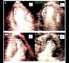
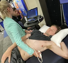
Cardiac ultrasound uses reflected sound waves (echos) to create images of anatomy inside the body. Echocardiograms are the primary cardiac imaging modality used to assess the heart and diagnose or track cardiac issues. Echo is the gold standard imaging modality to assess the heart, particularly with calculating left ventricular ejection fraction (LVEF), which is a measure of cardiac output. In addition to noninvasive standard transthoracic echo (TTE), invasive transesophgeal echo (TEE) is also used when clearer, more detailed imaging of the heart is needed. Both 3D and 4D echo echo systems are rapidly gaining wider adoption and enable new types of assessments, especially in the structural heart space and in transcatheter procedural guidance. Find news on general ultrasound imaging.

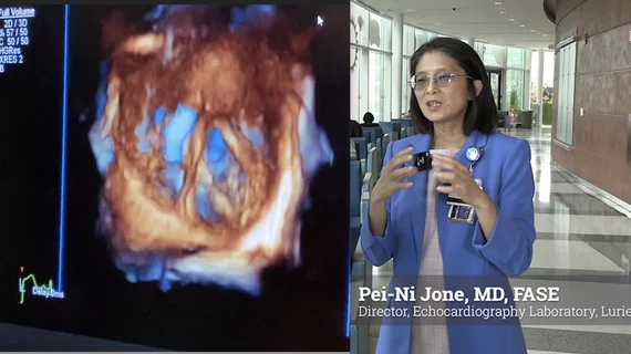
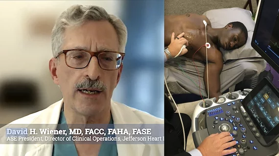
Video of ASE lobbyists explaining issues with Medicare funding policy reform in Washington DC.

Key government policies impacting cardiac ultrasound

Two experienced lobbyists who represent the American Society of Echocardiography detailed some of the biggest issues they are working on in Washington.

August 21, 2023
Video of Rebecca Hahn, MD, explains the roles and economics of interventional echocardiographers. #ASE #ASE23 #Structuralheart #echofirst

Interventional echocardiographers play a key role in the treatment of structural heart disease

Rebecca Hahn, MD, examines the growing role of the dedicated interventional echocardiographer and how these imagers are paid.

August 17, 2023
Video of Partho Sengupta, MD, explaining two major shifts in AI for echocardiography at ASE 2023. These include GPT and deep learning to automate measurements. #ASE23 #ASE2023 #ASE #AI

2 key reasons the use of AI in echocardiography is growing

Partho Sengupta, MD, an expert on the use of AI in cardiology, says some of the technologies being developed today will fundamentally change how cardiologists and sonographers do their jobs.

August 16, 2023
HCM expert Sherif F. Nagueh, MD, FACC, FASE, professor of cardiology, Methodist DeBakey Heart and Vascular Center, explains the key points of HCM imaging. #ASE #HCM

How medical imaging helps cardiologists treat hypertrophic cardiomyopathy

Hypertrophic cardiomyopathy expert Sherif F. Nagueh, MD, examined imaging's role in treating this serious condition. 

August 9, 2023
Photo of a surgical cardiac septal myectomy to treat hypertrophic cardiomyopathy (HCM). Nicholas Smedira MD discusses septal myectomy in HCM patients and the need for more detailed protocols for HCM care. #HCM #ASE #ASE2023 #myectomy

A surgeon's perspective on treating hypertrophic cardiomyopathy

Nicholas Smedira, MD, a cardiovascular surgeon with Cleveland Clinic, discusses septal myectomy and the need to improve overall HCM care with new standards and evidence-based guidelines.

August 9, 2023
Hari Naidu, MD, explains that inconsistent diagnostic imaging and how it is reported has likely led to a major underdiagnosis of HCM. #ASE #ASE2023 #HCM

Why hypertrophic cardiomyopathy may be widely underdiagnosed

One expert believes the lack of standardization for how diagnostic imaging in conducted and reported has led to a major underdiagnosis of HCM.

August 8, 2023
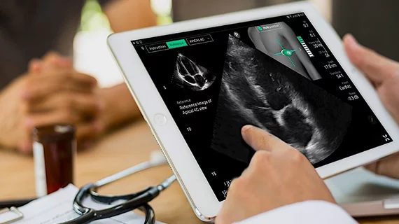
Ultrasight's AI to guide echo cardiac ultrasound POCUS exams at ASE 2023. The thumbnail of the chest shows the user how to the position the transducer and where to move or stop for image acquisition. The bar graph shows the quality of the image.

FDA clears new AI to guide point-of-care cardiac ultrasound exams

The FDA approved UltraSight's AI to show ultrasound users how to perform a diagnostic quality cardiac ultrasound exam at the point of care in multiple settings.

July 31, 2023
Matt Martinez, MD, Morristown Medical Center HCM program, explains how hypertrophic cardiomyopathy patient management is changing.

Exercise plays a key part in HCM management

Matthew Martinez, MD, detailed the many ways HCM care management is changing during an exclusive interview at ASE 2023.

July 26, 2023