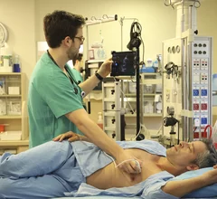
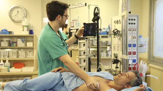
Echocardiography

Cardiac ultrasound uses reflected sound waves (echos) to create images of anatomy inside the body. Echocardiograms are the primary cardiac imaging modality used to assess the heart and diagnose or track cardiac issues. Echo is the gold standard imaging modality to assess the heart, particularly with calculating left ventricular ejection fraction (LVEF), which is a measure of cardiac output. In addition to noninvasive standard transthoracic echo (TTE), invasive transesophgeal echo (TEE) is also used when clearer, more detailed imaging of the heart is needed. Both 3D and 4D echo echo systems are rapidly gaining wider adoption and enable new types of assessments, especially in the structural heart space and in transcatheter procedural guidance. Find news on general ultrasound imaging.
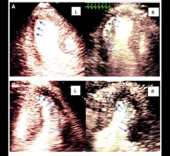
Intravascular imaging-guided PCI linked to improved survival, fewer adverse events in large new analysis

The study’s authors tracked data from nearly 16,000 patients who underwent PCI from 2010 to 2023. This is believed this to be the largest comparison of intravascular imaging-guided PCI and angiography-guided PCI to date.

Luc Mertens, MD, section head of echocardiography at The Hospital for Sick Children in Toronto, explains application of ultrafast ultrasound and blood speckle imaging in echocardiography

Vector flow and blood speckle echocardiography imaging may unveil new clinical insights

Luc Mertens, MD, Hospital for Sick Children, Toronto, explains research and potential uses for ultrafast echo imaging.

Staff at the new Fetal Cardiac Clinic at the Heart Institute at Children's Hospital Los Angeles. From left, Director of the Heart Institute David Romberger, RN, MSN, CCRN; attending physician Shuo Wang, MD; Executive Director of the Heart Institute Jennifer Klunder, MHA; Director of the Fetal Cardiac Clinic Jodie Votava-Smith, MD; Chief of the Division of Cardiology and Co-director of the Heart Institute Paul F. Kantor, MBBCh, MSc, FRCPC; and congenital cardiac surgeon Luke Wiggins, MD.

Children’s Hospital Los Angeles unveils new fetal cardiac clinic

Clinic for expectant mothers designed to better diagnose and treat congenital heart defects and heart diseases before birth. 

AI cardiology heart artificial intelligence deep learning

AI could be a game-changer for TAVR, but cardiologists remain ‘irreplaceable’

The rise of TAVR as a go-to treatment option for many AS patients has been one of the biggest stories in cardiology for several years now. How will advanced AI models impact this trend going forward? 

Video interview with Marielle Scherrer-Crosbie, MD, Hospital of the University of Pennsylvania, who explains details of creating a cardio-oncology program, whoi should be involved and the role of cardiac imaging.

Key things to remember when creating a cardio-oncology program

Marielle Scherrer-Crosbie, MD, explains what is needed to create a cardio-oncology program, and the role played by cardiac imagers.

Marielle Scherrer-Crosbie, MD, Hospital of the University of Pennsylvania, discusses the latest trends in cardiac strain echo.

Interest in strain echo imaging is rising, but hurdles remain

Marielle Scherrer-Crosbie, MD, told Cardiovascular Business that the current lack of reimbursement for strain echocardiography has been a challenge for care teams. 

transthoracic echocardiogram images from 2024 ASE guidelines

American Society of Echocardiography shares updated expert recommendations on pediatric, neonatal echo

These new guidelines provide a fresh look at topics ASE has not addressed with official society recommendations in many years. 

Royal Philips X11-4t Mini 3D TEE transducer

FDA clears smaller 3D TEE transducer for imaging children, high-risk adult patients

The newly approved device is much smaller than previous offerings, helping operators capture 3D images during structural heart evaluations.