Echocardiography

Cardiac ultrasound uses reflected sound waves (echos) to create images of anatomy inside the body. Echocardiograms are the primary cardiac imaging modality used to assess the heart and diagnose or track cardiac issues. Echo is the gold standard imaging modality to assess the heart, particularly with calculating left ventricular ejection fraction (LVEF), which is a measure of cardiac output. In addition to noninvasive standard transthoracic echo (TTE), invasive transesophgeal echo (TEE) is also used when clearer, more detailed imaging of the heart is needed. Both 3D and 4D echo echo systems are rapidly gaining wider adoption and enable new types of assessments, especially in the structural heart space and in transcatheter procedural guidance. Find news on general ultrasound imaging.

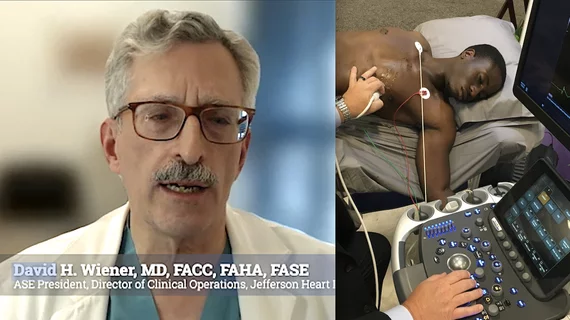
Ritu Thamman, MD, FASE, University of Pittsburgh School of Medicine, discusses trends and the technical aspects of strain imaging. #ASE #ASE23 #ASE2023 #strainecho

Strain imaging making a significant impact on the treatment of heart patients

Strain imaging has gained considerable momentum in recent years as an effective tool for getting more value out of heart evaluations. Ritu Thamman, MD, provided an in-depth look at the latest trends in strain echocardiography in an exclusive interview. 

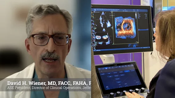
Video of Denise Garris, ASE regulatory consultant and principal of the Korris Group, explaining how reimbursements from insurance andMedicare work to pay or cardiac ultrasound exams. #ASE #ASE360 #ASE23 #ASE2023 #healthcarereimbursment

Understanding reimbursements and coding for echocardiography

"I think everyone would agree that the current fee-for-service model is not working appropriately and there needs to be a fix," regulatory consultant Denise Garris explained in a new interview. 

Steven Lester, MD, Mayo Clinic, explains how AI will soon revolutionize the echo lab. Photo by Dave Fornell. #ASE #ASE360

How AI can make echo labs more efficient

Advances in AI technology are expected to give physicians more time to provide high-quality care to their patients.

Arzhang Fallahi, MD, and David Hsi, MD, discussing imaging-based aortic stenosis screening

Q&A: Cardiologists explore the potential impact of a screening program for aortic stenosis

We already screen patients for breast cancer and lung cancer on a regular basis. Why not establish screening programs for aortic stenosis?

Jackie Sohn, DO, explains the use and trating requirements for intensivists and anesthesiologists to train on using point of care ultrasound (POCUS) at ASE 2023.

Training noncardiologists to perform basic POCUS exams

Jackie Sohn, DO, discussed how POCUS is now incorporated into fellowship training programs and the different ways these handheld and smaller cart-based systems are being utilized in hospitals.

Video of Juan Carlos Plana Gomez MD explaining how to create a cardio-oncology program at ASE 2023. #ASE #ASE23 #ASE2023

How to create a cardio-oncology program

Juan Carlos Plana Gomez, MD, discussed how cardiology and oncology programs can work together to provide high-quality patient care.

Purvi Parwani, MD, director of echocardiography, Loma Linda University Medical Center, explains the trend where heart failure imaging guidelines are driving a rising use of mixed multimodality imaging. #ASE #ASE2023

Multimodality imaging helps cardiologists manage heart failure patients—with an assist from AI

Purvi Parwani, MD, discussed the trend toward multimodality imaging for heart failure management. All modalities have their own weaknesses, she explained. 

Example of the Siemens Origin AI-automated cardiac ultrasound system performing auto contours and measurements after the AI sees what is being imaged and the operator hits the AI button on the console. The system is designed to be an assistant to the operator and knows the next steps in the exams.

Siemens Healthineers unveils 'truly revolutionary' AI-enabled echo system

The new-look system is able to evaluate a situation and anticipate what users may need next.