Computed Tomography

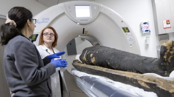
Computed tomography (CT) is a fast and accurate imaging modality often used in emergency settings and trauma imaging. CT scans, with or without (or both) iodinated contrast are frequently used to image the brain, chest, abdomen and pelvis, but also have post-imaging reconstructive capabilities for detailed orthopedic imaging. It is now a standard imaging modality in emergency rooms to quickly assess patients. CT uses a series of X-ray images shot as the gantry rotates around the patient. Computer technology assembles these into into a dataset volume than can be slices on any access, or advanced visualization software can extract specific parts of the anatomy for study. Find more content specific to cardiac CT.

lung cancer screening

More assertive language in cancer screening reports leads to likelier receipt of follow-up care

Patients with Lung-RADS 4A findings who spoke with a clinician by phone were three times more likely to see things through, experts wrote in JAMA Network Open

MRA for pulmonary embolus

MR angiography a suitable alternative to CT when ruling out pulmonary embolus

The modality switch became especially important during the iodinated contrast shortage of 2022 when clinics were forced to deploy mitigation tactics as a means of preserving their contrast supply. 

Thumbnail

Photon-counting technology offers new opportunities in imaging high-risk CAD patients

Coronary CT angiography is commonly used to assess patients at low or intermediate risk of CAD but is less effective in high-risk patients with an increased presence of coronary calcifications and stents due to the “blooming” effect they have on imaging.

quality

American College of Radiology tells CMS new quality measure would pose too much burden

The agency is proposing a new electronic clinical quality measure for inpatients tied to "excessive radiation dose or inadequate image quality." 

COVID causes increase risk of clotting. Example of a pulmonary embolism. CT pulmonary angiograms in a 77-year-old man with COVID-19 and a saddle embolus to pulmonary arteries (black arrow in A) extending into right and left pulmonary arteries (white arrows) in (A, B) axial and (C) coronal planes. Arrowheads show pulmonary changes associated with COVID-19 and possible lung infarction (black arrow in C). (D) Axial image at the level of the ventricles shows right-sided heart strain. RSNA. COVID PE

'Concerning' CT findings may lead to unnecessary hospitalization in PE patients

CT findings that have historically been deemed as concerning relative to PE might not hold as much weight when it comes to associated risks as previously thought, new analysis suggests.

Thumbnail

AI primed for pulmonary nodule detection in adults, falls short in pediatric population

Artificial intelligence tools have proven to be beneficial in detecting pulmonary nodules on chest CTs of adults, but less is known about their utility in pediatric populations. 

Prompting primary care physicians in the EHR helps boost use of CT for lung cancer screening

Rutgers University has seen significant increases in the number of fully filled out patient records, individuals eligible for screening, and completed LDCT orders. 

Radiologist-recommended testing for primary hyperparathyroidism improves diagnosis

Biochemical evaluation for patients with incidentally detected enlarged parathyroid glands can prompt earlier diagnosis, according to new data in the American Journal of Roentgenology