Computed Tomography

Computed tomography (CT) is a fast and accurate imaging modality often used in emergency settings and trauma imaging. CT scans, with or without (or both) iodinated contrast are frequently used to image the brain, chest, abdomen and pelvis, but also have post-imaging reconstructive capabilities for detailed orthopedic imaging. It is now a standard imaging modality in emergency rooms to quickly assess patients. CT uses a series of X-ray images shot as the gantry rotates around the patient. Computer technology assembles these into into a dataset volume than can be slices on any access, or advanced visualization software can extract specific parts of the anatomy for study. Find more content specific to cardiac CT.

Contrast protocols based on lean body weight save supplies, reduce patient risk

Adjusting contrast dose based on a patient’s lean body weight provides the same image quality as other administration methods, new data show.

Thumbnail

Former smokers who vape now still need to be screened for lung cancer. Many of them aren't

Despite kicking their initial habit, these individuals remain at risk of developing lung cancer due to their history of smoking cigarettes, current use of e-cigarettes, or both, experts caution.

Thumbnail

Radiologists, thoracic surgeons urge CMS to make millions more eligible for lung cancer screening

Enacting the requested modifications would make about 5 million more Americans eligible for LCS while saving about 21% more lives. 

Thumbnail

New study calls ED imaging protocols for geriatric head injuries into question

The new data suggest that admitting patients for observation and additional imaging to monitor for delayed intracranial hemorrhage might often be unnecessary.  

The feasibility of reduced contrast flow rates in PE studies

Around one out of every 1,000 people in the U.S. are diagnosed with a pulmonary embolism every year, but even more undergo imaging to rule out clots.  

Peter Monteleone, MD, an interventional cardiologist, national director of cardiovascular research at Ascension Health, and assistant professor, UT Austin Dell School of Medicine, explained the use of artificial intelligence (AI) to independently identify an emergency stroke or pulmonary embolism (PE) finding on a CT scan and automatically alert critical care team members. His health system uses this type of AI for earlier activation of the pulmonary embolism response team (PERT).

AI critical care software revolutionizes emergency response

Ascension Health in Texas uses AI that can read CT scans for stroke and pulmonary embolism to activate care teams before the images even get into the PACS.

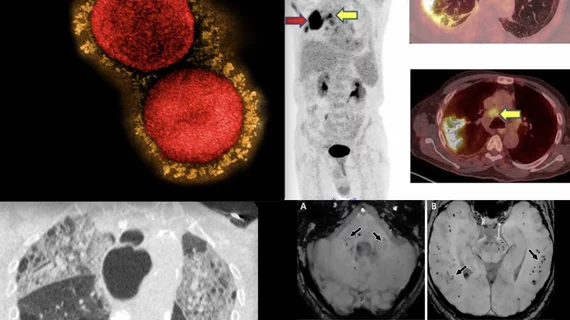
Imaging study reveals 'significant deformities' in lungs of some COVID patients

These changes could have long-lasting implications for lung function, authors of the new study warn.

lung cancer screening

New data underline importance of expanding Medicaid to address poor lung cancer screening uptake

“Also needed are institutional and societal efforts to address LCS capacity, including screening infrastructure investments and quality benchmarking," experts wrote in JAMA Internal Medicine