Magnetic Resonance Imaging

Cardiac magnetic resonance imaging MRI is used as both a functional and anatomical cardiac imaging test. It offers excellent soft tissue detail and the ability to quantify cardiac function. MRI scans can be performed with or without gadolinium contrast depending on what information is needed. Unlike computed tomography (CT), MRI does not use X-ray radiation, but patients with metal implants may have contraindications for MRI use because MR will heat up most metal objects. MRI exams usually take much longer than CT scans. How does MRI work? MR creates images by using powerful magnets to polarize hydrogen atoms in water (the body is made of of more than 80% water) so they face in one direction. A radiofrequency pulse is then used to ping these atoms, causing them to wobble, or resonate. The MRI coils detect this and computers can assemble images from the signals. Basic MRI scans will focus on the resonance of fat and water in two different sequences, which highlight and contrast different features in the anatomy.


COVID-19 vaccine associated myocarditis on short-axis 1.5T MRI images of a 19-year-old man who presented with chest pain three days following the second dose of an mRNA COVID-19 vaccine.

New cardiac MRI analysis offers updated insight into long-term impact of vaccine-related myocarditis

Months after their initial myocarditis diagnosis, late gadolinium enhancement (LGE) was observed in 62% of patients on follow-up cardiac MRI.

Thumbnail

Water-fat separation sequence yields superior image quality compared to standard coronary MRA

The 3-T Dixon GRE magnetic resonance angiography method produced better image quality and yielded greater overall diagnostic performance, according to a new study in AJR.

New-look imaging technique could change the way we see blood vessels

The new method, still being tested with mouse models, could help clinicians see blood vessels with improved clarity. 

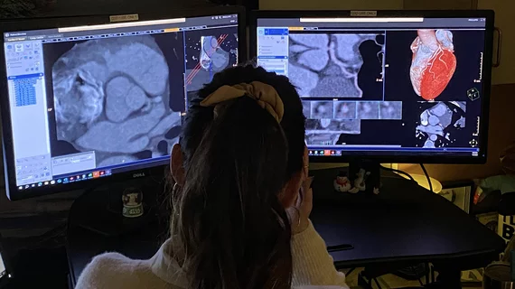
A dedicated Cardiac MRI scanner. The presence of myocardial fibrosis on cardiac MRI can help anticipate adverse arrhythmic events and  may offer a way to improve patient selection for cardiac implantable electronic devices (CIEDs). This is a dedicated cardiac MRI system at Baylor Scott White.

AI tool detects CVD on cardiac MRI in 20 seconds with high precision

The algorithm is already in use at some institutions in Europe, with plans for it to be utilized globally at many more within the year.

COVID-19 vaccine associated myocarditis on short-axis 1.5T MRI images of a 19-year-old man who presented with chest pain three days following the second dose of an mRNA COVID-19 vaccine.

Cardiac MRI sheds new light on vaccine-related myocarditis

Vaccine-associated myocarditis shows an injury pattern on cardiac MRI similar to other causes of myocarditis, but abnormalities are less severe. 

TEER associated with an added benefit for SMR patients in earlier stages of heart failure

Low levels of residual mitral regurgitation after TEER can make a big impact on patient outcomes, researchers explained in a new analysis. 

Early CMR imaging improves diagnosis of unexplained heart attacks

Turning to the modality just a few days earlier appeared to make a significant impact when it came to making an accurate diagnosis. 

Patients with non-MR compatible cardiac devices, including pacemakers and ICDs, can safely undergo MRI exams

"We had no issues with any of the patients and no harm to the devices," one researcher reported. The analysis included more than 500 patients.