Magnetic Resonance Imaging

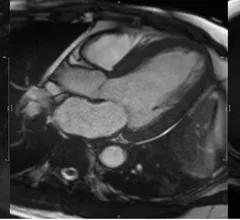
Cardiac magnetic resonance imaging MRI is used as both a functional and anatomical cardiac imaging test. It offers excellent soft tissue detail and the ability to quantify cardiac function. MRI scans can be performed with or without gadolinium contrast depending on what information is needed. Unlike computed tomography (CT), MRI does not use X-ray radiation, but patients with metal implants may have contraindications for MRI use because MR will heat up most metal objects. MRI exams usually take much longer than CT scans. How does MRI work? MR creates images by using powerful magnets to polarize hydrogen atoms in water (the body is made of of more than 80% water) so they face in one direction. A radiofrequency pulse is then used to ping these atoms, causing them to wobble, or resonate. The MRI coils detect this and computers can assemble images from the signals. Basic MRI scans will focus on the resonance of fat and water in two different sequences, which highlight and contrast different features in the anatomy.


women doctors

Cardiologist compensation hits $622K, up 32% from 10 years ago

According to a new analysis from MedAxiom, an American College of Cardiology company, cardiologist compensation has nearly returned to where it was before the COVID-19 pandemic. The detailed report also examined new patient visits, cardiac imaging utilization and much more. 

September 15, 2022

Cardiac MRI reveals that even competitive athletes are subject to reduced cardiac function after COVID

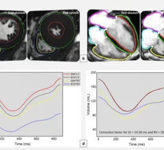
Compared to healthy controls, the COVID group displayed significantly worse global and segmental circumferential systolic strain.

August 15, 2022
This latest shortage involves dobutamine, an injectable medication often prescribed for advanced heart failure and cardiogenic shock. It is also a central component of dobutamine stress echocardiography (DSE), a test used to evaluate patients with severe aortic stenosis.

A new supply issue hits cardiology: 5 recommendations for cardiologists during the dobutamine shortage

The shortage could lead to significant issues for cardiologists. The American College of Cardiology provided some words of wisdom on the ongoing issue.  

August 3, 2022
CMR of a patient showing evidence of myocardial hypertrophy

Cardiac MRI findings linked with worse outcomes in hypertension patients

Nonischemic LGE was revealed to be independently associated with worse outcomes in hypertension patients. 

July 21, 2022

“We need an answer now”: Cardiologist awarded $2.3M to study cannabis use and heart health in people living with HIV

The four-year analysis will use cardiac MRI scans to closely monitor the heart health of people living with HIV who regularly use cannabis to treat their symptoms. 

July 6, 2022
SNMMI Image of the Year 68Ga-FAPI-46 PET/CT heart attack acute myocardial infarction

‘Image of the Year’ highlights the predictive power of a new PET imaging agent

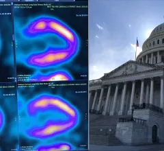
The Society of Nuclear Medicine and Molecular Imaging has chosen its 2022 Image of the Year, and it’s one that is sure to interest anyone in the field of cardiac imaging.

June 15, 2022
heart_body_blood_bloodvessel.jpg

Cardiac MRI findings can predict incident CVD years before onset

Prior imaging of the descending thoracic and abdominal aorta in study participants revealed thoracic aortic wall area (AWA), plaque prevalence and plaque volumes to be independently associated with incident CVD.

June 1, 2022
inHEART Medical, an international healthcare company with offices in France and the United States, has received clearance from the U.S. Food and Drug Administration (FDA) for its new 3D visualization software designed to improve the management of ventricular tachycardia (VT) ablation and other cardiac ablation procedures.

3D cardiac modeling solution for VT ablation receives FDA clearance

The new software solution was designed with ventricular tachycardia ablation and other cardiac ablation procedures in mind.

May 24, 2022