Magnetic Resonance Imaging

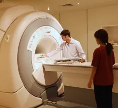
Cardiac magnetic resonance imaging MRI is used as both a functional and anatomical cardiac imaging test. It offers excellent soft tissue detail and the ability to quantify cardiac function. MRI scans can be performed with or without gadolinium contrast depending on what information is needed. Unlike computed tomography (CT), MRI does not use X-ray radiation, but patients with metal implants may have contraindications for MRI use because MR will heat up most metal objects. MRI exams usually take much longer than CT scans. How does MRI work? MR creates images by using powerful magnets to polarize hydrogen atoms in water (the body is made of of more than 80% water) so they face in one direction. A radiofrequency pulse is then used to ping these atoms, causing them to wobble, or resonate. The MRI coils detect this and computers can assemble images from the signals. Basic MRI scans will focus on the resonance of fat and water in two different sequences, which highlight and contrast different features in the anatomy.

T2 mapping may uncover cardiotoxic marker early enough to prevent heart failure

T2 mapping derived from weekly cardiac MRIs helped researchers identify cardiotoxicity at an early and reversible stage, a finding which may have implications for cancer patients at risk of chemotherapy-induced heart failure.

February 21, 2019

New protocol cuts costs, time for CMR imaging

A simplified cardiac magnetic resonance (CMR) protocol using a contrast agent allowed clinicians in Peru to diagnose heart ailments more quickly and cheaply, a new study found. The results, published online Aug. 29 in the Journal of the American Heart Association, highlight a potential pathway to realizing the benefits of CMR in developing nations.

August 29, 2018

Quantitative CMR improves predictions for patients with stable CAD

A quantitative measure of stress perfusion by cardiac magnetic resonance (CMR) imaging showed superior ability to predict major adverse cardiac events (MACE) in unselected patients with suspected coronary artery disease (CAD), according to a study published May 7 in JACC: Cardiovascular Imaging.

May 9, 2018

Image reconstruction algorithm, MRI-derived heart strain values can aid prognosis in amyloidosis patients

Recent research found strain parameters taken from a cine MRI-based deformable registration algorithm (DRA) can determine the severity of amyloid buildup in the heart and may provide prognostic information on patients with light-chain (AL) amyloidosis.

April 24, 2018

Seeing the Big Picture: Training Today’s Imagers to ‘Think Multimodality’

Cardiologists are receiving more exposure to different imaging modalities during their fellowships, but their job prospects and training vary widely. A more comprehensive and multimodality training approach could lead to better results.  

January 18, 2018