Echocardiography

Cardiac ultrasound uses reflected sound waves (echos) to create images of anatomy inside the body. Echocardiograms are the primary cardiac imaging modality used to assess the heart and diagnose or track cardiac issues. Echo is the gold standard imaging modality to assess the heart, particularly with calculating left ventricular ejection fraction (LVEF), which is a measure of cardiac output. In addition to noninvasive standard transthoracic echo (TTE), invasive transesophgeal echo (TEE) is also used when clearer, more detailed imaging of the heart is needed. Both 3D and 4D echo echo systems are rapidly gaining wider adoption and enable new types of assessments, especially in the structural heart space and in transcatheter procedural guidance. Find news on general ultrasound imaging.

Dr. Nee Khoo, director of the echocardiography lab, Stollery Children’s Hospital, Edmonton, Canada, explains the rapidly growing adoption of pediatric 3D transesophageal echo (TEE) to not only preplan, but also guide and assess the results of congenital heart surgeries.

The push to get 3D echo into the congenital operating room

Nee Khoo, MBChB, director of the echocardiography lab at Canada's Stollery Children’s Hospital, explains the rapid adoption of pediatric 3D transesophageal echo.

ASE leading push to revise CPT code for interventional echocardiography

The pressure is on for ASE and other societies to create a new CPT code for the use of interventional echocardiography during structural heart procedures.

As cancer therapies become increasingly targeted and complex, the need for sophisticated cardiovascular monitoring has grown in parallel. Cardiovascular Business spoke with Daniel Addison, MD, director of the cardio-oncology program at The Ohio State University and chair of the American Heart Association’s (AHA) Cardiac Imaging Committee, who said the use of multimodality imaging is transforming how clinicians manage cardiovascular risk in cancer patients and survivors.

Multimodality imaging is more important than ever to the field of cardio-oncology

“Multimodality imaging in cardio-oncology is something many of us in the field are truly excited about," Daniel Addison, MD, told Cardiovascular Business. He noted that modalities other than echocardiography are starting to play a much larger role in the treatment of these patients.

SCAI ICE position statement

SCAI highlights the benefits of ICE-guided structural heart interventions

ICE is gaining momentum as a helpful imaging option during structural heart interventions, according to a new SCAI position statement. TEE and TTE remain incredibly important, but there are times when ICE can provide the guidance necessary.

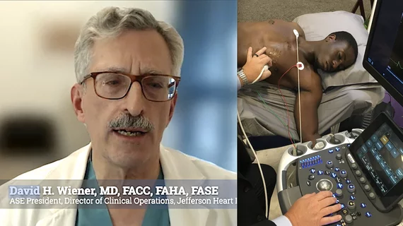
The co-chairs of the American Society of Echocardiography (ASE) 2025 meeting explain highlights from the in-person sonographer training sessions and focused sessions on difficult episodes of care.

ASE 2025 delivered a fully immersive experience for cardiovascular ultrasound professionals

The co-chairs of the American Society of Echocardiography's 2025 meeting wanted to "highlight the unique value of being in person." They looked back at the three-day event with Cardiovascular Business.

Cardiologists have developed an open-source artificial intelligence (AI) model capable of accurately evaluating a long list of measurements on echocardiography results. The group detailed the development and validation of that model in the Journal of the American College of Cardiology.

Open-source AI model delivers accurate echocardiography assessments

The algorithm, trained on more than 150,000 TTE studies, can calculate 18 different measurements regularly used in echocardiography

Exploring the link between left ventricular diastolic function and AFib

Evaluating LVDF with echocardiography or AI-powered electrocardiography can help identify individuals at an increased risk of developing atrial fibrillation, according to new data presented at the ASE 36th Annual Scientific Sessions.

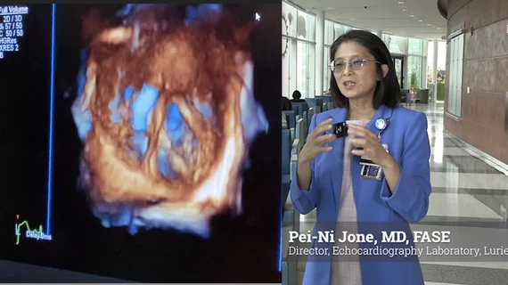
Pei-Ni Jone, MD, FASE, director, echocardiography laboratory, Lurie Children’s Hospital Heart Center, professor of pediatric cardiology, Northwestern University Feinberg School of Medicine, member of the American Society of Echocardiography (ASE) Board, and the chair of the ASE Pediatric and Congenital Heart Disease Council Steering Committee, explains how 3D echo is being used to better plan, guide and followup in congenital cardiac surgery.

3D echo guidance seeing increasing use in congenital heart surgery

Pei-Ni Jone, MD, FASE, director, echocardiography laboratory, Lurie Children’s Hospital Heart Center, member of the American Society of Echocardiography (ASE) Board, explains how 3D echo is being used to better plan, guide and follow up in congenital cardiac surgery.