Artificial Intelligence

Artificial intelligence (AI) is becoming a crucial component of healthcare to help augment physicians and make them more efficient. In medical imaging, it is helping radiologists more efficiently manage PACS worklists, enable structured reporting, auto detect injuries and diseases, and to pull in relevant prior exams and patient data. In cardiology, AI is helping automate tasks and measurements on imaging and in reporting systems, guides novice echo users to improve imaging and accuracy, and can risk stratify patients. AI includes deep learning algorithms, machine learning, computer-aided detection (CAD) systems, and convolutional neural networks. 

Thumbnail

AI helps NIH researchers evaluate stem cell-derived tissues

Researchers working for the U.S. government have used deep learning to evaluate stem cell-derived tissue samples, sharing their findings in the Journal of Clinical Investigation.

Thumbnail

3D neural network can help radiologists ID scarring associated with deadly heart condition

The boost in efficiency for measuring such scarring could make it easier for clinicians to overcome the time-consuming process of quantifying late gadolinium enhancement (LGE)—a proven predictor of hypertrophic cardiomyopathy.

Thumbnail

XACT Robotics announces $36M financing round

XACT Robotics, a radiology technology company with offices in the United States and Israel, raised $36 million in its latest financing round.

Thumbnail

Researchers worry that AI could be scaring students away from radiology

That’s according to a new survey of healthcare stakeholders, highlighted in November’s European Journal of Radiology

Thumbnail

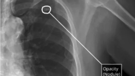
AI helps radiologists spot lung cancer on chest x-rays

In fact, clinicians who took a second look at x-rays using the deep learning software improved their sensitivity, on average, by 5.2%.

Thumbnail

Neural network improves imaging technique for an advanced look at cancer cells

The novel method uses a deep neural network to improve fluorescence lifetime imaging, which allowed scientists at Rensselaer Polytechnic Institute to view molecular-level interactions within cells.

Thumbnail

Researchers use color x-ray scanner, ‘GPS particles’ to pinpoint microfractures

Besides pinpointing microfractures, the researchers believe combining color spectral CT imaging with their novel nanoparticles could help detect more serious problems such as heart blockages.

Thumbnail

7T MRI reveals new view of damage in multiple sclerosis patients

"The 7T MRI scanner affords us new ways of viewing areas of damage in neurologic diseases such as MS that were not well seen using 3T MRI," corresponding author Jonathan Zurawski, MD, said.