Ultrasound

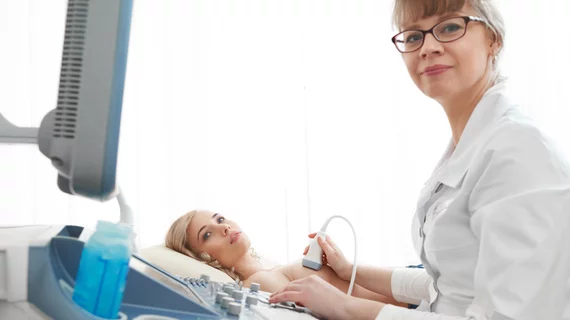
Ultrasound, also referred to as sonography or diagnostic ultrasound, uses high-frequency sound waves to visualize soft tissue. Ultrasounds are frequently ordered to measure fetal anatomy during pregnancy, check for blood clots and to guide needle biopsy procedures of the breast, abdomen and pelvis. The imaging modality does not use any radiation to create images. Find news specific to cardiac ultrasound (echocardiography).

Artificial intelligence automated measurements on an echocardiogram on the Siemens SyngoDynamics cardiovascular imaging and information solution. AI is helping speed workflows and complete tedious tasks faster and more accurately that humans, allowing sonographers and cardiologists to be more efficient. Photo by Dave Fornell

AI technologies to be featured heavily at ASE 2023

Artificial intelligence will be one of the hottest topics at the upcoming American Society of Echocardiography meeting in National Harbor, Maryland. 

Video of Jon Lindner explaining the use of ultrasound and bubble contrast for therapy.

Cardiac ultrasound could lead to key advances in heart attack care, drug delivery

Jonathan Lindner, MD, offers an update on the use of echocardiography and bubble contrast agents in a therapy role to help revascularize STEMI patients and increase drug and gene delivery.

Thumbnail

Prostate cancer size is frequently underestimated on imaging

Newly published data recently revealed just how common the extent of prostate cancer is underestimated based on imaging measurements. 

Considerations for supplemental imaging should extend beyond breast density, analysis suggests

Women with increased breast density are targeted significantly more for supplemental imaging than those without dense tissue, but there are other factors that increase the risk of mammography screening failure that should also be considered, according to new data.

Thumbnail

Ultrasound AI could help standardize trauma care

Trauma patients who present with poor blood flow and suspected abdominal hemorrhage are well served by emergency physicians using AI-augmented FAST imaging.

Katie Couric on USPSTF recommendations on when women should start getting mammograms

'Kind of a bummer': Katie Couric disappointed in USPSTF recs that exclude supplementary imaging for dense breasts

The task force cited insufficient evidence as the driver behind the decision to forego recommending supplemental screening. 

Thumbnail

RadNet, cancer care advocates praise passage of bills to boost imaging coverage

The policy changes forbid insurers from saddling patients with out-of-pocket costs for services associated with lung and breast cancer.

X-ray photon trajectory during the simulation phantom study from the side and top views. Due to scattering of the X-rays when they hit the lower end of the patient bed, exposure in mainly to the lower body of the interventional echocardiographer performing transesophageal echocardiography. The green lines are the scattered photon trajectories calculated by Monte Carlo simulation in the study.

Radiation exposure in the cath lab: Tracking the impact on interventional echocardiographers

Researchers found that echocardiographers in the cath lab are exposed to high doses of radiation on the right half of their body, especially the waist and lower body.