Ultrasound

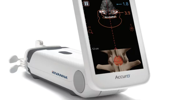
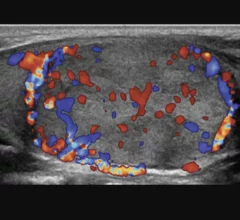
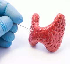
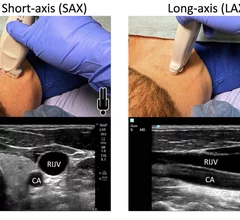
Ultrasound, also referred to as sonography or diagnostic ultrasound, uses high-frequency sound waves to visualize soft tissue. Ultrasounds are frequently ordered to measure fetal anatomy during pregnancy, check for blood clots and to guide needle biopsy procedures of the breast, abdomen and pelvis. The imaging modality does not use any radiation to create images. Find news specific to cardiac ultrasound (echocardiography).

An international panel of experts recently developed and validated a reporting assessment scoring system that analyzes the location and extent of prostate cancer recurrence. 

Prostate cancer size is frequently underestimated on imaging

Newly published data recently revealed just how common the extent of prostate cancer is underestimated based on imaging measurements. 

June 12, 2023
ultrasound.jpg

Considerations for supplemental imaging should extend beyond breast density, analysis suggests

Women with increased breast density are targeted significantly more for supplemental imaging than those without dense tissue, but there are other factors that increase the risk of mammography screening failure that should also be considered, according to new data.

June 12, 2023
Ambulance911

Ultrasound AI could help standardize trauma care

Trauma patients who present with poor blood flow and suspected abdominal hemorrhage are well served by emergency physicians using AI-augmented FAST imaging.

May 31, 2023
Katie Couric on USPSTF recommendations on when women should start getting mammograms

'Kind of a bummer': Katie Couric disappointed in USPSTF recs that exclude supplementary imaging for dense breasts

The task force cited insufficient evidence as the driver behind the decision to forego recommending supplemental screening. 

May 11, 2023

RadNet, cancer care advocates praise passage of bills to boost imaging coverage

The policy changes forbid insurers from saddling patients with out-of-pocket costs for services associated with lung and breast cancer.

May 5, 2023
X-ray photon trajectory during the simulation phantom study from the side and top views. Due to scattering of the X-rays when they hit the lower end of the patient bed, exposure in mainly to the lower body of the interventional echocardiographer performing transesophageal echocardiography. The green lines are the scattered photon trajectories calculated by Monte Carlo simulation in the study.

Radiation exposure in the cath lab: Tracking the impact on interventional echocardiographers

Researchers found that echocardiographers in the cath lab are exposed to high doses of radiation on the right half of their body, especially the waist and lower body. 

April 28, 2023
Example of interventional echocardiography TEE imaging superimposed on live fluoro during a transseptal puncture for a MitraClip procedure.

New ASE guideline outlines training standards for interventional echocardiography

The American Society of Echocardiography (ASE) released a new guideline document that outlines uniform training standards for interventional sonographers guiding structural heart procedures.

April 26, 2023
shear-wave elastography TIRADS

Shear wave elastography assessment improves TI-RADS scoring

That’s according to an award-winning scientific online poster presented this week during the American Roentgen Ray Society’s annual meeting being held in Honolulu, Hawaii.

April 20, 2023