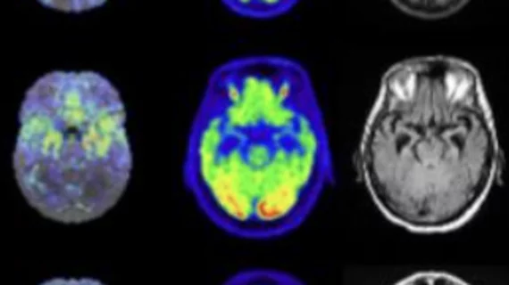
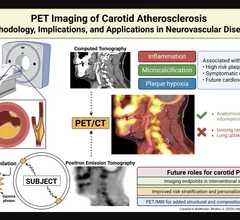
Positron emission tomography/computed tomography is a hybrid nuclear medicine imaging technique that helps radiologists spot abnormal metabolic activity. PET/CT is commonly used to diagnose cancers, heart diseases and certain brain disorders, among other conditions.
Currently, the organization relies on a mobile PET unit that parks once every week. However, demand has grown for PET/CT in the area, rising by around 60% over the last three years alone.
Limbic-predominant age-related TDP-43 encephalopathy, also known as LATE, was recently recognized as a type of dementia that occurs in older adults. It typically presents as memory-related cognitive decline.
Amyloid-related imaging abnormality edema is a known side effect of anti-amyloid monoclonal antibody treatments, such as Leqembi and donanemab Kisunla.
The move will make the Huntsman Cancer Institute the first academic medical center in the U.S. to produce Posluma, according to Blue Earth Diagnostics.
One of the tracer’s more significant advantages is its extended half-life of 109 minutes—significantly longer than other currently available PET MPI imaging agents.
The exam effectively helps providers manage patients’ treatment options, but for some, the costs associated with those treatments may negate the value of its findings.
Targeting CXCR4 during PET scans could help providers gain vital information regarding patients' potential to fully recover from myocardial infarction.
Beyond amyloidosis, nuclear cardiology is also increasingly used to image inflammation and infection in the myocardium, especially in diseases such as cardiac sarcoidosis and myocarditis.
PET has already gained significant momentum in recent years. Now, researchers are pointing to another possible use for the popular imaging modality: assessing carotid artery atherosclerosis.
GLP-1 receptor agonists alter glucose metabolism and the movement of food through the digestive system and can lead to unique uptake patterns on FDG PET-CT.