Molecular Imaging

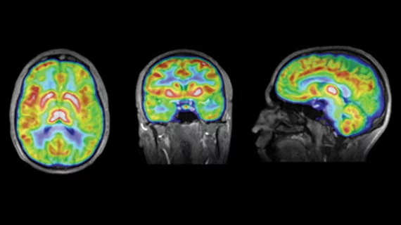
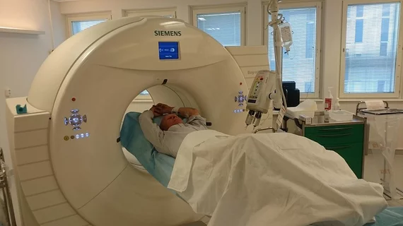
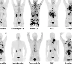
Molecular imaging (also called nuclear medicine or nuclear imaging) can image the function of cells inside the body at the molecular level. This includes the imaging modalities of positron emission computed tomography (PET) and single photon emission computed tomography (SPECT) imaging. How does PET and SPECT imaging work? Small amounts of radioactive material (radiopharmaceuticals) injected into a patient. These can use sugars or chemical traits to bond to specific cells. The radioactive material is taken up by cells that consume the sugars. The radiation emitted from inside the body is detected by photon detectors outside the body. Computers take the data to assemble images of the radiation emissions. Nuclear images may appear fuzzy or ghostly rather than the sharper resolution from MRI and CT.  But, it provides metabolic information at a cellular level, showing if there are defects in the function of the heart, areas of very high metabolic activity associated with cancer cells, or areas of inflammation, data not available from other modalities. These noninvasive imaging exams are used to diagnose cancer, heart disease, Alzheimer’s and Parkinson’s disease, bone disorders and other disorders. 

Thumbnail

Medical isotope update: Shortage expected to last weeks as nuclear reactor remains sidelined

Mo-99, Lu-177, and I-131 supply issues are forecasted until approximately one week after the downed reactor resumes operations on Feb. 12, the Nuclear Medicine Europe Emergency Response Team said.

Thumbnail

Nuclear reactor in Poland steps up to address Mo-99 shortage

The Maria reactor in Poland began producing the isotope hours after a water leak shut down the high flux reactor in Petter, the Netherlands.

Thumbnail

Medical isotope shortage looms as 'unplanned' outage halts Mo-99, Lu-177 production

The root cause of the problem has not yet been discovered, halting isotope production for an undisclosed amount of time, according to SNMMI.

Thumbnail

PSMA-PET validates commonly used system measuring risk of prostate cancer recurrence

"The accuracy of PSMA-PET is essential to improve stratification and potentially outcomes both in low-risk and high-risk settings,” a doctor involved in the study explained in the Journal of Nuclear Medicine.

SNMMI images of unusual imaging pattern observed on FDG PET/CT or FDG PET/MR that may be due to Omicron COVID-19 infection. Unlike the FDG PET/CT pattern seen with infections from previous strains of COVID-19, with principal involvement of the lungs, this new array of findings is primarily centered in the upper aerodigestive tract and cervical lymph nodes. What does omnicron COVID look like in medical imaging?

Unusual pattern on PET/CT may indicate COVID omicron variant

The Society of Nuclear Medicine and Molecular Imaging offered up a handful of tips to help providers who encounter such findings.

Thumbnail

FDA approves Telix’s prostate cancer imaging product, improving access to PSMA-PET

The company said it can provide its toolkit for preparing gallium-68 to more than 85% of eligible PET imaging sites within its large network of U.S. nuclear pharmacies.

Thumbnail

PSMA PET/CT better detects prostate cancer spread, long-term outcomes over conventional imaging

While the results offer indirect evidence, UCLA researchers said they support the clinical use of this technique for initial patient assessment.

Texas A&M University to lead isotope R&D trainee program with $2 million grant

Of the 17 institutions collaborating on the project, eight are minority-serving organizations.