Magnetic Resonance Imaging

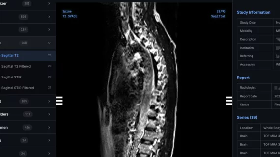
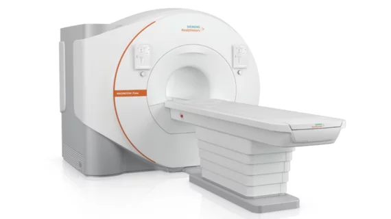
Magnetic resonance imaging (MRI) is the gold standard imaging modality for soft tissues. It produces detail cross-sectional images of soft tissue and bone anatomy, including muscles, tendons, ligaments, brain and organs, without the use of ionizing radiation. In addition to orthopedic imaging, MRI is also used for heart, brain and breast. MRI uses gadolinium contrast in many exams to highlight tissues and blood vessels, which enhances images and offers better diagnostic quality. It can also be used in conjunction with PET scans. How does MRI work? MR creates images by using powerful magnets to polarize hydrogen atoms in water (the body is made of of more than 80% water) so they face in one direction. A radiofrequency pulse is then used to ping these atoms, causing them to wobble, or resonate. The MRI coils detect this and computers can assemble images from the signals. Basic MRI scans will focus on the resonance of fat and water in two different sequences, which highlight and contrast different features in the anatomy.

Thumbnail

Paris tech company scores FDA clearance for brain MRI assistant

QyScore helps clinicians pinpoint MRI markers, which signal the early onset of conditions such as Alzheimer's and Parkinson's. 

Thumbnail

AI helps bolster radiologists’ ability to detect ADHD using MRI

This breakthrough could help providers pinpoint other neurological conditions, experts noted in a study published Wednesday, Dec. 11, in Radiology: Artificial Intelligence. 

Thumbnail

Key brain region smaller in women who use birth control pills, MRI scans reveal

Women who use oral contraceptives have a “significantly” smaller area of the brain that’s responsible for regulating everything from body temperature to mood, appetite, sex drive and sleep cycles. 

Thumbnail

Doc notches key win in court battle over certificate of need for MRI

Gajendra Singh, MD, has been working for years to open up more options for consumers in the Winston-Salem area, but he reached a roadblock last year.

Thumbnail

Use of CT and MRI to diagnose eye problems in the ED skyrocketing, underlining need for guidance

Johns Hopkins researchers recently made that determination by scouring data from millions of ED visits conducted between 2007 and 2015. 

Thumbnail

Opioid exposure in womb alters infants’ brain function, MRI scans reveal

Indiana University School of Medicine recently made that discovery using resting state functional MRI to scan the brains of 16 sleeping newborns. 

Thumbnail

How one hospital system is using patient-centered referral forms to reduce unnecessary CT and MRI scans

The inappropriate use of imaging tests to assess headaches and lower back pain is an ongoing issue in Canada, one that’s contributing to ballooning care costs and long wait times. 

Thumbnail

Harvard researchers use powerful 7T MRI to fill in details, root out progression of MS

"The 7T MRI scanner affords us new ways of viewing areas of damage in neurologic diseases such as MS that were not well seen using 3T MRI; it's capturing nuances that we would otherwise miss," said study co-author Jonathan Zurawski.