Magnetic Resonance Imaging

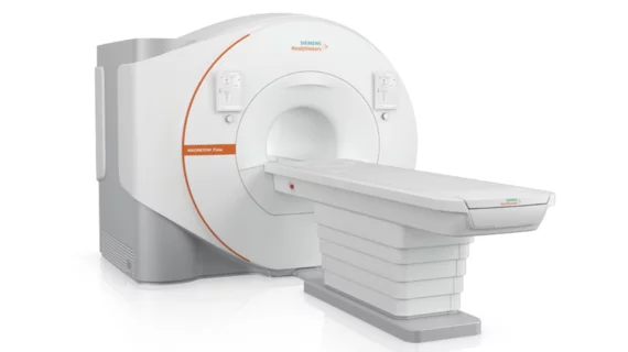
Magnetic resonance imaging (MRI) is the gold standard imaging modality for soft tissues. It produces detail cross-sectional images of soft tissue and bone anatomy, including muscles, tendons, ligaments, brain and organs, without the use of ionizing radiation. In addition to orthopedic imaging, MRI is also used for heart, brain and breast. MRI uses gadolinium contrast in many exams to highlight tissues and blood vessels, which enhances images and offers better diagnostic quality. It can also be used in conjunction with PET scans. How does MRI work? MR creates images by using powerful magnets to polarize hydrogen atoms in water (the body is made of of more than 80% water) so they face in one direction. A radiofrequency pulse is then used to ping these atoms, causing them to wobble, or resonate. The MRI coils detect this and computers can assemble images from the signals. Basic MRI scans will focus on the resonance of fat and water in two different sequences, which highlight and contrast different features in the anatomy.

Smart Choice MRI

Former executives of now-defunct Smart Choice MRI eye expansion with new imaging center chain

Atlanta-based ImageLink already has 14 locations across Georgia and Ohio, while a 15th just recently opened in Boynton Beach, Florida. 

spine lumbar

Standardizing MRI spine degradation definitions bolsters neuro, musculoskeletal radiologist agreement

Substantial variability in interpretation exists between rad subspecialties and spine clinicians, experts detailed in Current Problems in Diagnostic Radiology.

Supplemental, short-protocol MRI a cost-effective addition for stroke patients in the ED

Head CT can sometimes miss the early signs of minor strokes, German scientists wrote in European Radiology

Thumbnail

Contrast-enhanced mammography as effective as MRI at evaluating newly diagnosed breast cancer

CESM utilizing iodine offers a sound alternative to magnetic resonance, illuminating lesions otherwise invisible on a regular mammogram. 

Thumbnail

Abdominal MRI intervention helps drop respiratory motion and image quality degradation

Language barriers can pose problems in the MRI suite, where abdominal exams come with carefully choreographed breathing instructions, NYU experts wrote in JACR

pain pills opioids

Structural MRI shows promise predicting relapse among patients battling opioid addiction

The experimental diagnostic method combines imaging and machine learning to seek patterns in functional connectivity and brain structure data. 

prostate cancer PSA

Annual MRI surveillance of low-risk prostate cancer drops biopsies while preserving life expectancy

NYU imaging experts created a simulation model to determine the most cost-effective method of monitoring the disease. 

fatty liver disease hepatic steatosis

Liver MR imaging specialist eyes $75M initial public offering on the Nasdaq

Founded in 2012, Perspectum's flagship FDA-cleared software LiverMultiScan aids in the detection of disease using scans of the organ.