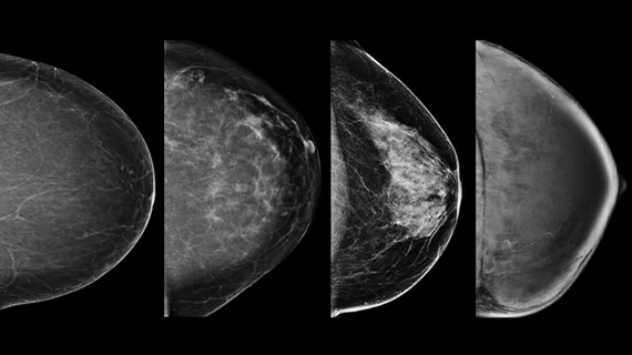
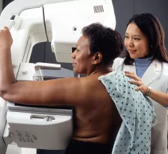
Diagnostic screening programs help catch cancer, abnormalities or other diseases before they reach an advanced stage, saving lives and healthcare costs. Screening programs include, lung, breast, prostate, and cervical cancer, among many others.
There are no standards requiring radiologists to report on the presence of BACs, even though up to half of referring providers have indicated they would prefer to be made aware of the finding.
Breast artery calcifications are already visible when radiologists review mammograms, but nothing typically happens with them. Researchers aimed to see if AI could help translate those findings into an easy-to-understand cardiovascular risk score.
Despite the great progress that has been made toward the clinical implementation of AI, new data caution against trusting the technology as a single reader in certain settings.
Nearly one million patients per year in the United States require treatment for plantar fasciitis, according to authors of a new study published in Academic Radiology.
The researchers looked at 73 eligible studies consisting of 3,930 malignant and 2,861 benign lesions that evaluated the diagnostic performance of DWI, diffusion-tensor imaging (DTI) and intravoxel incoherent motion (IVIM) in the breast.
CT colonography (CTC) achieved a similar positive predictive value (PPV) as colonoscopy during colon cancer screening and can help clinicians care for patients by specifying the nature of exam findings, according to a new study published in the American Journal of Roentgenology.
Rectal cancer patients given a “good prognosis” on MRI may be able to avoid preoperative chemoradiotherapy (CRT), a technique commonly associated with long-term adverse outcomes, according to results of a new study published in JAMA Oncology.
Combining digital breast tomosynthesis (DBT) with digital mammography (DM) can spot additional lesions in patients with breast cancer, reported authors of a recent study published in Radiology. However, the combined method did have limitations.
The American College of Physicians (ACP) now recommends women undergo mammography every other year, beginning at age 50. The new guidelines have drawn criticism from the American College of Radiology and Society of Breast Imaging.
Researchers have established normal ranges of bone density in a part of the lumbar spine that is routinely imaged incidentally. Their primary aim is to equip radiologists with data that can be referred to when reading chest and abdominal CTs so the reader can opportunistically cross-screen for osteoporosis and check for compression fractures.