Computed Tomography

Computed tomography (CT) is a fast and accurate imaging modality often used in emergency settings and trauma imaging. CT scans, with or without (or both) iodinated contrast are frequently used to image the brain, chest, abdomen and pelvis, but also have post-imaging reconstructive capabilities for detailed orthopedic imaging. It is now a standard imaging modality in emergency rooms to quickly assess patients. CT uses a series of X-ray images shot as the gantry rotates around the patient. Computer technology assembles these into into a dataset volume than can be slices on any access, or advanced visualization software can extract specific parts of the anatomy for study. Find more content specific to cardiac CT.

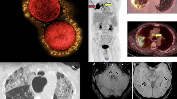
Multimodality imaging turns up serial thromboses following AstraZeneca COVID vaccination

Whole-body imaging reveals clinically undetectable blood clots in patients after their first dose of AstraZeneca's COVID vaccine.

Siemens debuts new, cleared SPECT/CT model

Siemens Healthineers splashed an FDA-approved SPECT/CT system June 12 at the 2022 annual meeting of the Society of Nuclear Medicine & Nuclear Imaging in Vancouver, B.C.

Thumbnail

Radiologists can reclaim an hour every day with AI assistance

The AI software assisted in various tasks, such as segmenting, labeling and measuring normal structures, providing an automated analysis of pulmonary, cardiac and musculoskeletal findings.

How COVID-19 made some radiologists newly nimble with workload management

During one surge, the hospital’s CT volumes spiked to 55% higher than pre-pandemic levels—no doubt due to heavy demand for advanced chest imaging.

CT scan showing lung cancer nodules with measurements of each nodule to track growth or regression from treatment. Image courtesy of RSNA

Downgrading lung nodules at 3-month follow-up 'may be problematic'

A new analysis suggests that follow-up imaging protocols for certain lung nodules identified via CT may need to be revisited. 

Predicting healthcare utilization in COPD patients using CT and machine learning

Combining CT lung measurements with machine learning models to predict prognosis in COPD patients could help to lessen their reliance on emergency services. 

rib fracture broken ribs

Advanced MSK imaging trio—CT, MR, ultrasound—justified for certain trauma patients upon incident and over time

The comprehensive approach may be warranted for patients whose musculoskeletal injuries include fractures of the costal cartilage. 

Brian Ghoshhajra, MD, MBA, division chief, cardiovascular imaging, Massachusetts General Hospital and associate professor of radiology, Harvard Medical School, and a board member of the Society of cardiovascular Computed Tomography (SCCT) explains the rapidly expanding interest in cardiac computed tomography (CT) under the new chest pain evaluation guidelines.

VIDEO: The new role of cardiac CT in chest pain evaluation

Brian Ghoshhajra, MD, MBA, division chief, cardiovascular imaging, Massachusetts General Hospital and associate professor of radiology, Harvard Medical School, and a board member of the Society of cardiovascular Computed Tomography (SCCT), explains the rapidly expanding interest in cardiac computed tomography (CT) under the new chest pain evaluation guidelines.