Computed Tomography

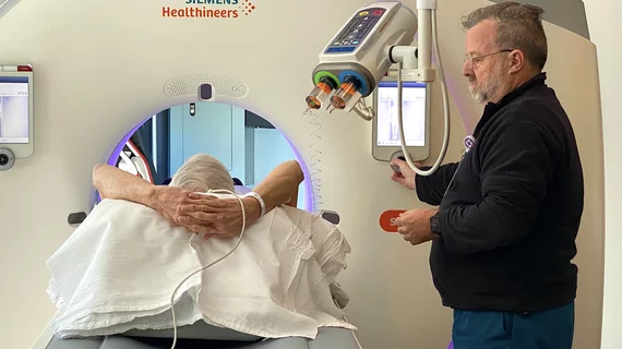
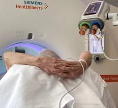
Computed tomography (CT) is a fast and accurate imaging modality often used in emergency settings and trauma imaging. CT scans, with or without (or both) iodinated contrast are frequently used to image the brain, chest, abdomen and pelvis, but also have post-imaging reconstructive capabilities for detailed orthopedic imaging. It is now a standard imaging modality in emergency rooms to quickly assess patients. CT uses a series of X-ray images shot as the gantry rotates around the patient. Computer technology assembles these into into a dataset volume than can be slices on any access, or advanced visualization software can extract specific parts of the anatomy for study. Find more content specific to cardiac CT.

Supply crunch forces ED to develop contrast use mitigation protocol for PE

The protocol led to a reduction in CT scans without sacrificing diagnostic accuracy.

cancer screening puzzle

World Health Organization forecasts 77% increase in new cancer cases by 2050

Two years ago, there were roughly 20 million cancer cases. However, population aging and growth are pushing these numbers upward, WHO said. 

Thumbnail

More with less: Department cuts technologists’ wasted time searching for CT supplies by 62%

The University of Utah Health department of radiology recently tackled a quality improvement project to quash wasted minutes spent seeking syringes and other items in storage. 

doors subspecialty specialize

Inconsistent imaging: Radiologists letting personal preferences influence decision-making

There is significant variation from one radiologist to the next in suggesting follow-up head and neck imaging, Harvard experts detailed in AJR

Kale Turtle Cook Museum of Natural Science

Rare sea turtle with infected shell undergoes CT scan

The procedure is medically necessary, but it also provides scientists insights into the Kemp’s ridley species of sea turtle.

Hip strain injury fracture broken hip

AI model predicts hip fractures in a short-term timeframe

Current risk assessments are unable to calculate the likelihood of a near-future fracture after a patient breaks their hip.

lung cancer screening

Patient-radiologist consultations, held in reading room after lung cancer screening, show promise

Conversations last 10 minutes or less, with specialists providing a preliminary interpretation of imaging findings in conjunction with smoking cessation counseling. 

Thumbnail

Study examines use of MSOT for detecting pancreatic cancer, improving surgery

The University of Oklahoma Health has earned a $3 million grant from the National Cancer Institute for this project.