Cardiac Imaging

While cardiac ultrasound is the widely used imaging modality for heart assessments, computed tomography (CT), magnetic resonance imaging (MRI) and nuclear imaging are also used and are often complimentary, each offering specific details about the heart other modalities cannot. For this reason the clinical question being asked often determines the imaging test that will be used.

Thumbnail

Is coronary heart disease on CT associated with early development of COPD?

"This awareness is important to avoid oversight of symptoms like dyspnea and vague chest discomfort, which can easily be interpreted as symptoms caused by the known disease COPD,” experts involved in the study said.

transcatheter aortic valve replacement (TAVR) procedure guided by CT fusion imaging on a patient with heart failure and a blood clot

Cardiologists and surgeons perform first TAVR using CT imaging guidance on HF patient with blood clot

“We believe this case is potentially groundbreaking," one interventional cardiologist said. 

The steps involved to gain transcaval access between the inferior vena cava and the aorta to avoid problematic anatomical areas that would have otherwise disqualified a patient for a femoral access TAVR procedure. Illustration of the NIH

Transcaval TAVR outperforms transaxillary TAVR when femoral access is not an option

Transcaval TAVR was linked to a much lower stroke rate and fewer bleeding complications. 

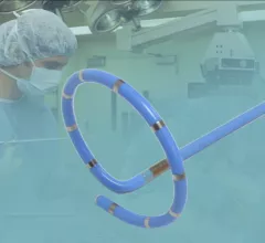
The Shockwave Medical M5+ new peripheral intravascular lithotripsy (IVL) catheter cuts procedures times in half and was showed for the first time at the American College of Cardiology (ACC) 2022 meeting. It is designed to break up calcium in heavily calcified arteries to avoid the need for vessel trauma caused by high pressure angioplasty.

VIDEO: Shockwave Medical demonstrates faster intravascular lithotripsy at ACC.22

The new Shockwave Medical M5+ peripheral intravascular lithotripsy (IVL) catheter made its debut at ACC.22. 

A dedicated Cardiac MRI scanner. The presence of myocardial fibrosis on cardiac MRI can help anticipate adverse arrhythmic events and  may offer a way to improve patient selection for cardiac implantable electronic devices (CIEDs). This is a dedicated cardiac MRI system at Baylor Scott White.

Could cardiac MRI become the reference standard for diagnosing heart failure?

Cardiac MRI may offer a more accurate and less invasive method for diagnosing heart failure, a new study published in the European Heart Journal shows. 

COVID-19 vaccine associated myocarditis on short-axis 1.5T MRI images of a 19-year-old man who presented with chest pain three days following the second dose of an mRNA COVID-19 vaccine.

New cardiac MRI analysis offers updated insight into long-term impact of vaccine-related myocarditis

Months after their initial myocarditis diagnosis, late gadolinium enhancement (LGE) was observed in 62% of patients on follow-up cardiac MRI.

Thumbnail

Cardiac implantable electronic devices can be safely reprogrammed to MRI-safe mode via remote operator

In 209 study participants, the remote reprogramming resulted in a successful, event-free MRI scan for every patient.

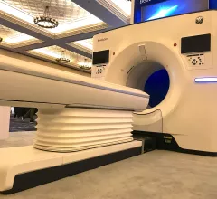
Siemens Healthineers showed examples at ACC 2022 of cardiac computed tomography (CT) from its new Naeotom Alpha photon-counting CT scanner cleared by the FDA in 2021.

VIDEO: Example of photo-counting cardiac CT with calcified coronaries

Siemens Healthineers showed examples at ACC 2022 of cardiac computed tomography (CT) from its new Naeotom Alpha photon-counting CT scanner cleared by the FDA in 2021.