Cardiac Imaging

While cardiac ultrasound is the widely used imaging modality for heart assessments, computed tomography (CT), magnetic resonance imaging (MRI) and nuclear imaging are also used and are often complimentary, each offering specific details about the heart other modalities cannot. For this reason the clinical question being asked often determines the imaging test that will be used.

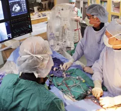
Interventional cardiology procedure in a cath lab at Beaumont Hospital.

PCI boosts survival for ischemic HF patients with moderate-to-severe mitral regurgitation

Mortality rates can be especially high for ischemic HF patients presenting with FMR and LVSD, but PCI may offer some relief. 

A CT image from the Heart Institute in Kyiv, Ukraine, showing a Russian bullet in a civilian patient's upper lung lobe. The patient was being treated at the cardiology hospital after he tried to drive his family out of the area of the front lines and was shot at by Russian soldiers. He is being treated by cardiac surgeon Igor Mokryk MD. Photo by Igor Mokryk.

Heart hospital in Ukraine treating wounded civilians

Cardiothoracic surgeon Igor Mokryk, MD, spent last week taking his family to the Polish border. This week, he treated his first gunshot wound patient at the Heart Institute in Kyiv.

chest pain lung pulmonary embolism

Image quality is not an issue for AI model that detects pulmonary embolisms on CT

CTPA is the standard of care for diagnosing PE, but suboptimal scans make it difficult to reach a diagnosis. A new Clinical Imaging study tests the effectiveness of AI when image quality is lacking.

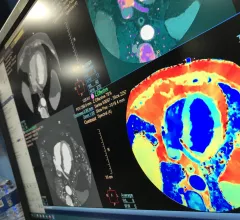
A dedicated Cardiac MRI scanner. The presence of myocardial fibrosis on cardiac MRI can help anticipate adverse arrhythmic events and  may offer a way to improve patient selection for cardiac implantable electronic devices (CIEDs). This is a dedicated cardiac MRI system at Baylor Scott White.

AI tool detects CVD on cardiac MRI in 20 seconds with high precision

The algorithm is already in use at some institutions in Europe, with plans for it to be utilized globally at many more within the year.

Thumbnail

What routine mammograms can tell us about a woman’s CVD risk

Signs of breast arterial calcification on a patient’s routine mammogram may suggest they face a greater risk of cardiovascular disease.

William A. Zoghbi, MD, MACC, FAHA, FASE, is the chair of the Department of Cardiology at the Houston Methodist DeBakey Heart and Vascular Center, and past president of both the American College of Cardiology (ACC) and the American Society of Echocardiography (ASE).

VIDEO: New advances in echocardiography

William A. Zoghbi, MD, past president of the ACC and ASE, discussed the latest trends in cardiac ultrasound technology. 

Thumbnail

AI-powered ECG analysis could boost care for patients with hypertrophic cardiomyopathy

Advanced algorithms can pick up on key details in a 12-lead ECG that human readers are unable to see. 

AFib, AI and heart-healthy diets: European Society of Cardiology previews EHRA 2022

The European Heart Rhythm Association's annual conference is headed to Denmark.