Computed Tomography

Cardiac computed tomography (CT) has become a primary cardiovascular imaging modality in the past 20 years, and was recommended as a 1A recommendation in the 2021 chest pain assessment guidelines. CT calcium scoring has became a primary risk assessment for coronary artery disease and whether patients should be on statins. Coronary CT angiography (CCTA) is used to for anatomical assessment of the arteries for plaque burden and to identify areas of blockage that may cause ischemia and heart attacks. Additional use of contrast CT perfusion or fractional flow reserve CT (FFR-CT) can offer physiological information on the function of the heart. CT plays a primary role in structural heart assessments for heart valves, repair of congenital defects and left atrial appendage occlusion (LAAO) for both pre-procedure planning and procedural guidance. Find more news on general radiology CT use.

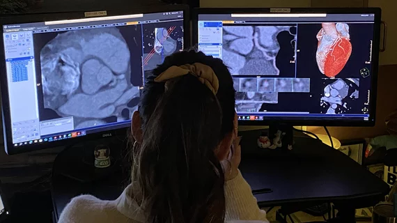
cardiologists evaluating the human heart to provide a treatment strategy

Managing stable angina: How cardiologists can find the right mix of revascularization, therapy and interventions

The management of stable angina has been evolving at a rapid rate. Using a one-size-fits-all strategy is becoming a thing of the past, replaced by a patient-centered approach that requires open communication and a healthy understanding of recent clinical research.

Ron Blankstein, MD, FACC, FASNC, MSCCT, FASPC, associate director, cardiovascular imaging program, director, cardiac computed tomography, Brigham and Women’s Hospital, and a professor of medicine and radiology, Harvard Medical School, explains a few of the recent advancements in cardiac MRI, nuclear imaging and computed tomography.

Exploring major breakthroughs in cardiac imaging

Ron Blankstein, MD, director of cardiac computed tomography for Brigham and Women’s Hospital, details recent developments in cardiac MRI, nuclear imaging and CT.

CMS may double Medicare payments for cardiac CT

The agency is gathering feedback on a proposal to double the amount hospitals are paid for coronary computed tomography angiography, with the comment period ending Sept. 9.

Echocardiography vs CT for Prediction of Newly Created LVOT Area during TMVR

3D echo comparable to CT for predicting TMVR complications

LVOT obstruction after TMVR remains a significant concern among structural heart cardiologists. Predicting the risk of LVOT obstruction typically requires CT, which comes with certain disadvantages compared to other imaging options.

The American Society of Nuclear Cardiology (ASNC) is asking Congress to repeal the appropriate use software provision mandate, which physicians say is an obstacle to efficient care.

ASNC asks Congress to officially repeal the AUC mandate for advanced medical imaging

Medicare rescinded the provision in the 2024 Medicare Physician Fee Schedule, but the law remains on the books. 

HeartFlow introduced its next generation artificial intelligence (AI) Plaque Analysis with an interactive experience at SCCT 2024. It shows a 3D plaque model and analysis by territory across calcified, non-calcified and low-attenuation plaques. This includes viewing cross-sectional, color-coded images of each plaque type where it was quantified along the vessel. #SCCT #SCCT24 #SCCT2024

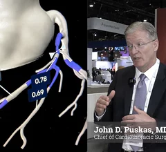
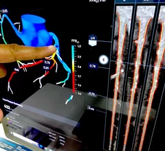
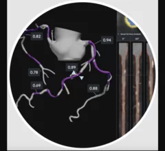
Updated HeartFlow technology offers interactive features, full integration with FFR-CT 

HeartFlow is using SCCT 2024 to introduce the world to its updated Plaque Analysis platform. “Accurately diagnosing a patient’s risk for coronary artery disease is critical for determining the best treatment," Chief Medical Officer Campbell Rogers, MD, explained.

Researchers with University College London and the European Synchrotron Radiation Facility (ESRF) have used a new X-ray technique, hierarchical phase-contrast tomography (HiP-CT), to capture images of the human heart in unprecedented detail. The group shared its images, as well as a full analysis, in Radiology.[1]

New imaging technique captures human heart with 'unprecedented detail'

The new images were captured at the European Synchrotron Radiation Facility using hierarchical phase-contrast tomography. One specialist called them "Google Earth for the human heart." 

SCCT aims to provide common language for CCTA use with updated guidance

The Society of Cardiovascular Computed Tomography has developed a new expert consensus document designed to get physicians on the same page when discussing this growing technology.