Computed Tomography

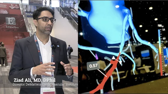
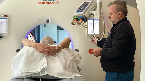
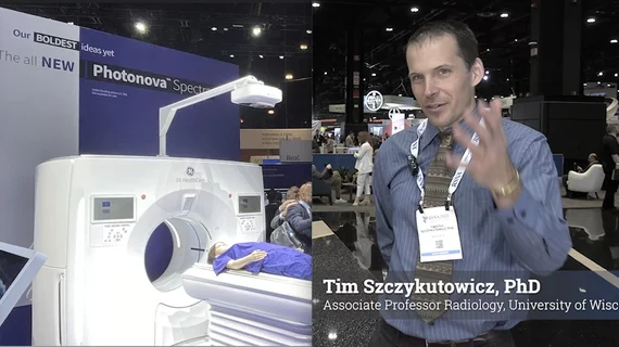
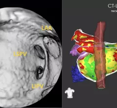
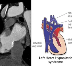
Cardiac computed tomography (CT) has become a primary cardiovascular imaging modality in the past 20 years, and was recommended as a 1A recommendation in the 2021 chest pain assessment guidelines. CT calcium scoring has became a primary risk assessment for coronary artery disease and whether patients should be on statins. Coronary CT angiography (CCTA) is used to for anatomical assessment of the arteries for plaque burden and to identify areas of blockage that may cause ischemia and heart attacks. Additional use of contrast CT perfusion or fractional flow reserve CT (FFR-CT) can offer physiological information on the function of the heart. CT plays a primary role in structural heart assessments for heart valves, repair of congenital defects and left atrial appendage occlusion (LAAO) for both pre-procedure planning and procedural guidance. Find more news on general radiology CT use.


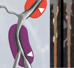
Perivascular fat attenuation index CT scans showing the amount coronary wall inflammation. This measure is being tested in trials to see if it can accurately predict which coronary artery lesions will progress to cause heart attacks.

Novel cardiac imaging biomarker could predict risk of coronary inflammation, heart attack

A team of international researchers has developed a new imaging biomarker able to non-invasively predict a patient’s risk of coronary inflammation and heart attack, according to research published Aug. 28 in The Lancet.

Han awarded the inaugural 2018 DeHaan Award for Innovation in Cardiology

Vienna, VA (August 13, 2018) — B. Kelly Han, MD, is the recipient of the inaugural 2018 DeHaan Award for Innovation in Cardiology, which includes a $200,000 grant.

HeartFlow Analysis Demonstrates Highest Diagnostic Performance for Detecting Heart Disease Compared to Other Non-Invasive Tests

PARIS, May 22, 2018 — HeartFlow, Inc. today announced new data from the PACIFIC trial showed that the HeartFlow Analysis had the highest diagnostic performance for detecting coronary artery disease (CAD), the most common form of heart disease, when compared to other commonly used non-invasive cardiac tests.

Thumbnail

Document offers guidance on curbing radiation exposure during CV imaging

An expert consensus document published May 2 in the Journal of the American College of Cardiology offers recommendations for limiting radiation exposure to both patients and medical personnel during cardiovascular imaging procedures.

Example of spectral, or dual-energy CT, confirming a pulmonary embolism (PE). Image courtesy of Philips Healthcare

MRA vs CTA for evaluating pulmonary embolism: Does the chosen modality impact downstream imaging utilization?

MR angiography (MRA) is a relatively new alternative to CT angiography (CTA) for the evaluation of suspected pulmonary embolism (PE) and it even has one major advantage over CTA: it does not expose patients to ionizing radiation.

Thumbnail

James K. Min named editor-in-chief of Journal of Cardiovascular Computed Tomography

James K. Min, MD, has been chosen to be the new editor-in-chief of the Journal of Cardiovascular Computed Tomography (JCCT), the official journal of the Society of Cardiovascular Computed Tomography (SCCT). He will serve a five-year term.
Thumbnail

Seeing the Big Picture: Training Today’s Imagers to ‘Think Multimodality’

Cardiologists are receiving more exposure to different imaging modalities during their fellowships, but their job prospects and training vary widely. A more comprehensive and multimodality training approach could lead to better results.  

Be Mindful of the Stark Law in CCTA Alliances

Numerous radiology groups are currently discussing arrangements whereby cardiologists and the radiologists would essentially split the responsibilities for reading coronary CT angiography (CCTA) studies. Most commonly, as proposed, the cardiologists would read and generate a signed interpretation report, but limited to the cardiac portion of the