Computed Tomography

Cardiac computed tomography (CT) has become a primary cardiovascular imaging modality in the past 20 years, and was recommended as a 1A recommendation in the 2021 chest pain assessment guidelines. CT calcium scoring has became a primary risk assessment for coronary artery disease and whether patients should be on statins. Coronary CT angiography (CCTA) is used to for anatomical assessment of the arteries for plaque burden and to identify areas of blockage that may cause ischemia and heart attacks. Additional use of contrast CT perfusion or fractional flow reserve CT (FFR-CT) can offer physiological information on the function of the heart. CT plays a primary role in structural heart assessments for heart valves, repair of congenital defects and left atrial appendage occlusion (LAAO) for both pre-procedure planning and procedural guidance. Find more news on general radiology CT use.

Leslee Shaw, PhD, MSCCT, FACC, MASNC, FAHA, director of the Blavatnik Family Women's Health Research Institute, a professor of medicine at the Icahn School of Medicine at Mount Sinai, and a former president of both SCCT and ASNC. She explains the sex differences in cardiovascular imaging presentations in women versus men.

VIDEO: CT can play a role in identifying women's differences in cardiovascular presentations

Leslee Shaw, PhD, director of the Blavatnik Family Women's Health Research Institute, a professor of medicine at the Icahn School of Medicine at Mount Sinai, and a former president of both SCCT and ASNC, explains the sex differences in cardiovascular imaging presentations in women versus men. 

Ricardo Cury, MD, MBA, MSCCT, chairman of radiology, direct of cardiac imaging, Baptist Health South Florida and Miami Cardiac and Vascular Institute, discusses the new CAD-RADS 2.0 cardiac imaging reporting criteria at the 2022 SCCT meeting. Interview with Radiology Business Editor Dave Fornell.

VIDEO: What is new with CAD-RADS 2.0 cardiac imaging reporting?

Ricardo Cury, MD, chairman of radiology and director of cardiac imaging, Baptist Health South Florida and Miami Cardiac and Vascular Institute, discusses the new CAD-RADS 2.0 cardiac imaging reporting criteria.

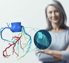
AI-generated coronary tree from a patient's CT scan showing a color code of areas of interest for plaque burden from the Cleerly software shown at SCCT 2022.

VIDEO: The role of AI in cardiac imaging

Ed Nicol, MD, president-elect of the Society of Cardiovascular Computed Tomography, provided us with an exclusive look at how AI is expected to change cardiac imaging.

Left, HeartFlow's RoadMap analysis enables cardiac CT readers to identify stenoses in the major coronary arteries. The AI provides visualization and quantification of the location and severity of anatomic narrowings. Right image, HeartFlow's Plaque Analysis AI algorithm automates assessment of coronary plaque characteristics and volume on CCTA exams to greatly reduce the time it takes to manually assess and quantify these features.

HeartFlow gains FDA clearance for 2 new AI-powered imaging assessments

The solutions, Plaque Analysis and RoadMap Analysis, both use coronary CT angiography to provide clinicians with a noninvasive look at patients who present with coronary artery disease and face a heightened myocardial infarction risk.

VIDEO: Cardiac CT as a front-line chest pain exam in the ED

Harold Litt, MD, explains the use of cardiac CT as a primary imaging exam for chest pain at his emergency department over the past decade.

CT has been used for planning and procedural guidance in pulmonary vein isolation (PVI) for about 20 years. It shows the anatomy to the electrophysiologist. The EPs also can load the imaging into their EP mapping system. It also shows them the location of the esophagus and the phrenic nerve so they can avoid these during the procedures. #PVI #EPeeps

VIDEO: The expanding role of cardiac CT in electrophysiology

Harold Litt, MD, division chief of cardiothoracic imaging and director of the Center for Advanced CT Imaging Sciences at the Perelman School of Medicine of the University of Pennsylvania, explains the role of cardiac CT in EP procedures.

New recommendations highlight the importance of CCTA when treating acute chest pain in the ED

The Society of Cardiovascular Computed Tomography developed and published the new document to help educate healthcare providers who regularly treat acute chest pain in the emergency department. 

CT imaging showing the congenital heart defect of left heart hypoplastic syndrome.

VIDEO: Congenital heart imaging with cardiac CT

As the population of patients with adult congenital heart disease grows, they are presenting to adult cardiology clinics and being imaged with CT. Many also do not have access or cannot be imaged by MRI, said Renee Bullock-Palmer, MD.