Reprieve Cardiovascular emerged from stealth mode this week with sizable financial backing and some big name board members to advance development for its intelligent, automated diuretic and fluid management system for heart failure.
A judge upheld a previous trial victory that cleared Prairie Cardiovascular Consults of alleged mishandling of a patient and not fully understanding the severity of the heart condition prior to their death.
The move will bring together 24 representatives—12 from each party—who will work to stimulate American AI innovation while mitigating AI-related threats.
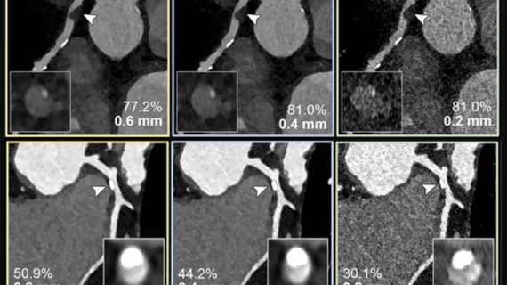
Examples of photon-counting coronary angiography showing how clarity improves as the thickness of the image is reduced. Top: 60-year-old female, with noncalcified plaque (arrowheads) and coronary stenosis (inset images). The reduced section thickness did not affect assessment in this patient. Bottom: 56-year-old female with calcified plaque (arrowheads) and coronary stenosis. The reduced section thickness leads to less calcium blooming and therefore a less severe percentage of stenosis. Photos courtesy of RSNA.
After a photon-counting CT, 54% of patients had their coronary artery disease classification downgraded.
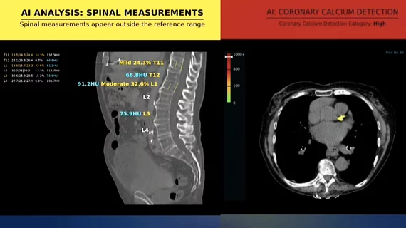
An example of an AI incidental opportunistic finding alert for coronary calcium in a CT scan unrelated to cardiology, detected using the Nanox AI algorithm.
Rads only reported this incidental finding in about 31% to 44% of cases, experts detailed in the Journal of the American College of Radiology.