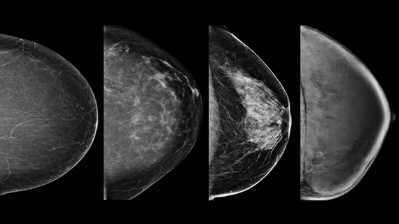
Example of the four types of breast tissue density. The density of fibroglandular tissue inside the breast impacts the ability to easily see cancers. On X-ray mammography, cancer and dense breast tissue both appear as white and can hide smaller cancers on 2D mammography. Dense breasts are also a risk factor for cancer. Cancers are very easy to spot in fatty breasts, but are almost impossible to find in extremely dense breasts. These examples show craniocaudal mammogram findings characterized as almost entirely fatty (far left), scattered areas of fibroglandular density (second from left), heterogeneously dense (second from right), and extremely dense (far right). Read more. Image courtesy of RSNA