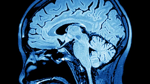
A: COVID-19-related spatial covariance pattern of cerebral glucose metabolism overlaid onto an MRI template. Voxels with negative region weights are color-coded in cool colors, and regions with positive region weights in hot colors. B: Association between the expression of COVID-19-related covariance pattern and the Montreal Cognitive Assessment (MoCA) score adjusted for years of education. Each dot represents a patient. C: Results of a statistical parametric mapping analysis. Upper row shows regions that show significant increases of normalized FDG uptake in COVID-19 patients at six-month follow-up compared to the subacute stage. Bottom row depicts regions that still show significant decreases of normalized FDG uptake in COVID-19 patients at six-month follow-up compared to the age-matched control cohort at an exploratory statistical threshold. Photo: G Blazhenets et al., Department of Nuclear Medicine, Medical Center – University of Freiburg, Faculty of Medicine, University of Freiburg.