Medical Imaging

Physicians utilize medical imaging to see inside the body to diagnose and treat patients. This includes computed tomography (CT), magnetic resonance imaging (MRI), X-ray, ultrasound, fluoroscopy, angiography,  and the nuclear imaging modalities of PET and SPECT. 

NRC petitioned by patient advocacy group to require reporting of extravasation

The group is advocating for the NRC to treat significant extravasations in the same manner as any other reportable medical event. 

DiA Imaging Analysis, which specialized in developing the AI-based automated cardiac ultrasound solution LVivo Seamless. The technology is now integrated through partnerships with dozens of healthcare vendors, including ScImage, GE Healthcare, Philips Healthcare Konica Minolta and IBM Watson.

ScImage latest vendor to adopt DiA Imaging Analysis AI for echocardiography

Artificial intelligence vendor DiA has emerged as a key third-party provider of AI to larger imaging vendors.

Thumbnail

Deep learning models predict COPD survival based on chest radiographs

Recently, experts explored the efficacy of two deep learning models for grading COPD severity based on chest radiographs—a method that has not yet been thoroughly tested.

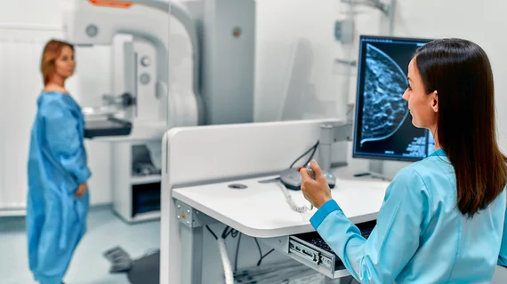
mammography mammogram breast cancer

‘You Only Look Once’ helps detect, classify lesions on deceptively normal screening mammograms

Researchers have combined three emerging technologies to detect and classify breast cancers found in follow-up imaging of women whose recent screening mammography was deemed normal.

Thumbnail

First ever full course of the latest MR-guided radiation therapy treatments completed at Henry Ford Health

The Detroit-based cancer institute achieved this landmark moment using the MRIdian A3i, which is the latest enhancement of the MRIdian system from ViewRay, Inc.

Brian Ghoshhajra, MD, MBA, division chief, cardiovascular imaging, Massachusetts General Hospital and associate professor of radiology, Harvard Medical School, and a board member of the Society of cardiovascular Computed Tomography (SCCT) explains the rapidly expanding interest in cardiac computed tomography (CT) under the new chest pain evaluation guidelines.

VIDEO: The new role of cardiac CT in chest pain evaluation

Brian Ghoshhajra, MD, MBA, division chief, cardiovascular imaging, Massachusetts General Hospital and associate professor of radiology, Harvard Medical School, and a board member of the Society of cardiovascular Computed Tomography (SCCT), explains the rapidly expanding interest in cardiac computed tomography (CT) under the new chest pain evaluation guidelines.

Radiologists best AI at diagnosing COVID on chest X-rays

AI can help humans inspect chest X-rays for COVID-19, but the technology is unfit to serve as a standalone diagnostic tool for that purpose. 

hip dysplasia joint socket

Diagnosing osteoporosis using a deep-radiomics-based approach

Researchers have developed deep-radiomics-based models capable of predicting osteoporosis with high accuracy and without need for DXA imaging.