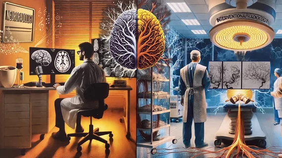
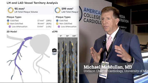
Medical Imaging

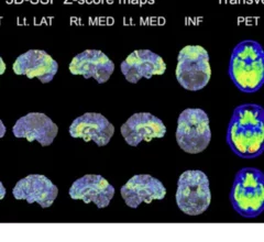
Physicians utilize medical imaging to see inside the body to diagnose and treat patients. This includes computed tomography (CT), magnetic resonance imaging (MRI), X-ray, ultrasound, fluoroscopy, angiography,  and the nuclear imaging modalities of PET and SPECT. 

Radiopharma firm Telix touts progress after FDA rejects brain imaging agent application

The Australia-based drug manufacturer is working to release Pixclara, a PET contrast agent for diagnosing glioma, a rare form of brain cancer. 

Ultrasound guidance a safer, more accurate method for biopsies of peripheral lung lesions in children

Ultrasound guidance safer, more effective for biopsies of peripheral lung lesions in pediatric patients

The technique has proven effective in adults, but until recently there has been a lack of data on its use among children.

breast ultrasound biopsy

New care approach cuts breast biopsy referrals by over 50%

Rising imaging volumes have led to an uptick in the unearthing of unexpected breast lesions, with a corresponding jump in follow-up examinations.

GE Healthcare's Venue Go point-of-care ultrasound (POCUS) system features AI-enabled tools to speed workflows, including Caption Guidance to help non-expert users capture diagnostic quality cardiac images. These systems helped drive positive earnings for the company in Q2 2025.

Bedside POCUS could save hospitals millions when used on patients with shortness of breath

“The explanation here is simple. Ultrasound gives you more information, and more concrete information, about what’s going on,” authors of a new JAMA study say.

GE HealthCare Revolution Vibe CCTA

FDA clears GE Revolution Vibe CT scanner aimed at rapidly growing CCTA market

“FDA clearance comes at the perfect time, with guidelines being adopted clinically for cardiac CT and with positive reimbursement driving procedure growth," says Jean-Luc Procaccini, CEO, molecular imaging and CT.

Radiologist to lead new research on blast injuries in service members

The study, which will be funded by a $2.3 million grant from the U.S. Department of Defense, will harness the power of advanced MRI technology to visualize the brain in a way that has not previously been possible.  

Pei-Ni Jone, MD, FASE, director, echocardiography laboratory, Lurie Children’s Hospital Heart Center, professor of pediatric cardiology, Northwestern University Feinberg School of Medicine, member of the American Society of Echocardiography (ASE) Board, and the chair of the ASE Pediatric and Congenital Heart Disease Council Steering Committee, explains how 3D echo is being used to better plan, guide and followup in congenital cardiac surgery.

3D echo guidance seeing increasing use in congenital heart surgery

Pei-Ni Jone, MD, FASE, director, echocardiography laboratory, Lurie Children’s Hospital Heart Center, member of the American Society of Echocardiography (ASE) Board, explains how 3D echo is being used to better plan, guide and follow up in congenital cardiac surgery.

Jaime Warren, EdD, FACC, vice president of care transformation at MedAxiom, an ACC company, explains what is needed to address staffing shortages created by a 94% increase in cardiac CT volumes over the past 5 years.

How to address CCTA staffing shortages

Jaime Warren, EdD, FACC, vice president of care transformation at MedAxiom, an ACC company, explains what is needed to address staffing shortages created by a 94% increase in cardiac CT volumes over the past 5 years.