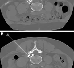
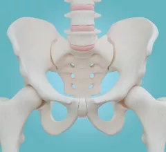
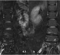
Orthopedic imaging relies on X-ray, MRI and CT to diagnose disorders and injuries affecting the bones, muscles, ligaments, tendons, cartilage, and spine. Orthopedists also use these test results to create an effective treatment plan.
Although gadolinium-based contrast agents are largely considered safe and are routinely used for MRI exams, experts suggest that providers should still utilize GBCAs sparingly for musculoskeletal studies.
In PRF procedures, a probe is used to intermittently apply energy directly to the dorsal root ganglia, which is often where pain and neurologic symptoms associated with sciatica originate.
Experts compared the cortical thickness of three bones—the radius, tibia and second metatarsal—of 200 volunteers who had undergone prior bone mineral density measurements to evaluate the ultrasound method’s accuracy.
The software was put to the test on nearly 5,000 trauma radiographs, assessing for the presence of fractures, dislocations, elbow effusions and focal bone lesions. In some cases, it outperformed radiologists by as many as 82 points.
Female authorship in musculoskeletal radiology research has seen a steady climb over the last two decades—a positive trend that is indicative of progress in a field where leadership is largely dominated by males.