Neuroimaging

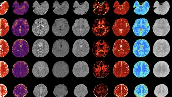
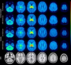
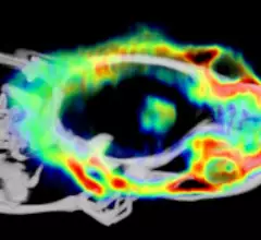
Neuroradiology is a branch of medical imaging focused on spotting abnormalities of the central and peripheral nervous system, spine, head and neck. These highly trained doctors use CT, MRI, X-ray and other techniques to diagnose strokes, tumors, aneurysms and other neurological conditions.

Thumbnail

'Significant' brain abnormalities shown to persist 6 months after COVID recovery

The regions that displayed the most significant differentiations in susceptibility values are known to be associated with neurological conditions such as fatigue, insomnia, anxiety, depression, headaches and other cognitive issues—all common complaints among many COVID long haulers. 

Thumbnail

Experts discover brain connectivity patterns unique to children with autism

The findings were also found to be associated with the severity of symptoms in individuals with ASD.

Thumbnail

New lesion measurement better predicts MS progression

Researchers recently presented a new method that considers how already existing lesions grow over time as an indicator of disease progression.

Thumbnail

New study may give insight into the neurologic dysfunction of some long-COVID patients

Vessel wall imaging—an MRI technique that offers high resolution images of the vessel wall—may hold clues as to the mechanisms by which COVID infections continue to cause neurologic symptoms after initial recovery in some patients.  

A convicted murderer's fate could hinge on different interpretations of her brain scans

In the case of a convicted murderer, a New York neuro specialist cited a slew of neuroimaging findings that indicate “severe dysfunction” that could have numbed the defendant’s “brakes of inhibition.” 

Thumbnail

How maternal COVID-related stress can alter fetal brain development

The preliminary results of a new study published in the medRxiv preprint server highlight variances in fetal brain development based on mothers’ perceived stress during the onset of COVID.

MRI shows structural, functional brain abnormalities in Lyme disease long-haulers

Compared to a group of patients who had not been previously diagnosed with Lyme disease, those who had been infected displayed unusual activity in the frontal lobe of the brain on functional MRI scans.

Structural brain abnormalities linked to functional seizures

Using MRI imaging, the experts found that patients who experienced functional seizures had thinning in the superior temporal cortex and thickening of the left occipital cortex.