Magnetic Resonance Imaging

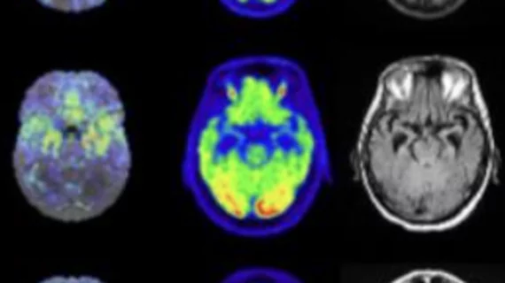
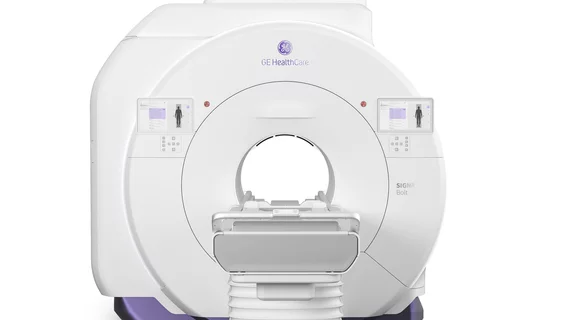
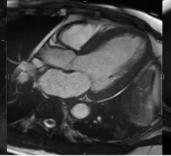
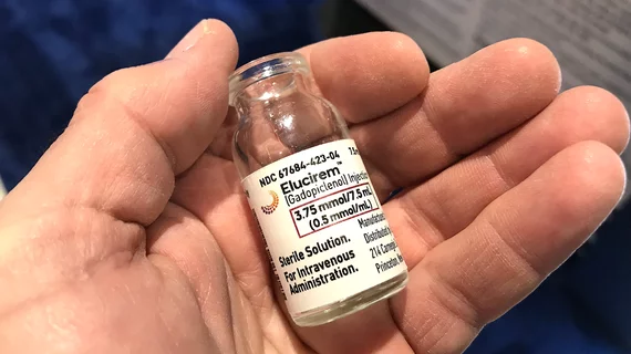
Magnetic resonance imaging (MRI) is the gold standard imaging modality for soft tissues. It produces detail cross-sectional images of soft tissue and bone anatomy, including muscles, tendons, ligaments, brain and organs, without the use of ionizing radiation. In addition to orthopedic imaging, MRI is also used for heart, brain and breast. MRI uses gadolinium contrast in many exams to highlight tissues and blood vessels, which enhances images and offers better diagnostic quality. It can also be used in conjunction with PET scans. How does MRI work? MR creates images by using powerful magnets to polarize hydrogen atoms in water (the body is made of of more than 80% water) so they face in one direction. A radiofrequency pulse is then used to ping these atoms, causing them to wobble, or resonate. The MRI coils detect this and computers can assemble images from the signals. Basic MRI scans will focus on the resonance of fat and water in two different sequences, which highlight and contrast different features in the anatomy.

Thumbnail

FDA clears tool for producing muscle assessment scores from MRI scans

AMRA Medical said its product uses a rapid neck-to-knee imaging protocol to produce actionable, patient-specific reports. 

Thumbnail

MRI study brings scientists closer to diagnosing CTE in living patients

"This is very exciting because it brings us that much closer to detecting CTE in living people," explained lead author of the study, Michael Alosco, PhD.

Multi-slice knee MRI technique saves time without sacrificing quality

Radiologists should know that the more they use simultaneous multi-slice settings, the more likely artifacts are to appear.

Thumbnail

Artificial intelligence could pinpoint success of rectal cancer treatment based on routine MRI

The Department of Defense is gifting Case Western researchers a grant to study the use of AI in determining whether patients require surgery. 

Goldberg MRI stroke brain

New CT protocol uses scout images to expedite stroke patients' path to MRI

The new safety screening protocol should "markedly improve" current delays in screening patients, Mayo Clinic doctors explained.

Brain MRI volumes dip but acuity leaps, with implications for radiologist staffing

More complicated exams could require additional physician time and effort, Emory University experts detailed in Current Problems in Diagnostic Radiology

Thumbnail

Prenatal MRI reveals ‘major’ brain differences among unborn babies exposed to alcohol

About 9.8% of women confess to drinking alcohol during their pregnancy, a number that's likely much higher, experts said Wednesday.

Thumbnail

COVID-19 infection in pregnant mothers does not harm developing fetuses’ brains

Radiology experts analyzed fetal MRIs from 33 women who contracted the virus while bearing a child, sharing their results at RSNA21.